Serviços Personalizados
Journal
Artigo
Indicadores
-
 Citado por SciELO
Links relacionados
-
 Similares em
SciELO
Compartilhar
Revista Pan-Amazônica de Saúde
versão impressa ISSN 2176-6215versão On-line ISSN 2176-6223
Rev Pan-Amaz Saude v.1 n.1 Ananindeua mar. 2010
http://dx.doi.org/10.5123/S2176-62232010000100021
ARTIGO ORIGINAL | ORIGINAL ARTICLE | ARTÍCULO ORIGINAL
Molecular characterization of norovirus, sapovirus and astrovirus in children with acute gastroenteritis from Belém, Pará, Brazil
Caracterização molecular de norovírus, sapovírus e astrovírus em crianças com gastroenterite aguda em Belém, Pará, Brasil
Caracterización molecular de norovirus, sapovirus y astrovirus en niños con gastroenteritis aguda en Belém (Pará, Brasil)
Glicélia Cruz Aragão; Darleise de Souza Oliveira; Mirleide Cordeiro dos Santos; Joana D'Arc Pereira Mascarenhas; Consuelo Silva de Oliveira; Alexandre da Costa Linhares; Yvone Benchimol Gabbay
Seção de Virologia, Instituto Evandro Chagas/SVS/MS, Ananindeua, Pará, Brasil
Endereço para correspondência
Correspondence
Dirección para correspondencia
ABSTRACT
The importance of norovirus (NoVs), sapovirus (SaVs) and human astrovirus (HAstVs) as causes of gastroenteritis outbreaks are already well-defined, but a few studies have described sporadic cases of acute gastroenteritis caused by these viral entities. The aim of this study was to determine the role of these viruses in the etiology of acute gastroenteritis in children enrolled to participate in hospital – and emergency department – based intensive surveillance carried out in Belém, Brazil, from March to September 2003. A total of 305 stool specimens from patients with severe gastroenteritis were collected and screened by reverse transcription followed by polymerase chain reaction (RT-PCR), using the specific primers Mon 269 and Mon 270 for HAstVs, p289 and p290 for human calicivirus (HuCVs), and Mon 431/433 and Mon 432/434 for NoVs. Sequencing of RT-PCR HAstV, HuCVs and NoVs amplicons was carried out using the same primers. Of the 305 samples tested, 96 (31.5%) were positive, with 51 diagnosed as HuCVs, 40 as HAstVs and five as mixed infections. Of the 56 (18.4%) HuCVs sequenced, 30 were NoVs (9.8%) of genogroups GI-4 and GII-4, and 15 (4.9%) were SaVs of types GI-1, GI-2 and GII-1. HAstVs, including genotypes 1, 8 and 2, were detected in 45 (14.7%) samples. This study has highlighted the importance of these viruses as causes of acute gastroenteritis and established the circulation of different genotypes during the study period. These results reinforce the need for establishing an intensive surveillance for gastroenteritis caused by these viruses to assess the burden of disease and to monitor the circulation of genotypes.
Keywords: Norovirus; Sapovirus; Human, Astrovirus; Gastroenteritis; Molecular Sequence Data.
RESUMO
A importância dos norovírus (NoVs), sapovírus (SaVs) e astrovírus humanos (HAstVs) como causa de surtos de gastroenteritis já está bem definida. Entretanto, poucos estudos têm descrito casos esporádicos de gastroenterites aguda causados por esses agentes. O objetivo deste estudo foi determinar o papel destes vírus na etiologia da gastroenterite aguda em crianças atendidas durante uma vigilância intensiva realizada em hospitais e ambulatórios de Belém, Brasil, de março a setembro de 2003. Um total de 305 espécimes fecais de pacientes com gastrenterite grave foram coletados e testados por reação em cadeia da polimerase precedida de transcrição reversa (RT-PCR), utilizando iniciadores específicos Mon 269 e Mon 270 para os HAstVs; p289 e p290 para os calicivírus humanos (HuCVs); e Mon 431/433 e Mon 432/434 para os NoVs. Sequenciamento dos amplicons de HAstV, HuCVs e NoVs, obtidos por RT-PCR, foi realizado usando os mesmos iniciadores. Das 305 amostras testadas, 96 (31,5%) apresentaram resultados positivos, sendo que 51 diagnosticadas como HuCVs, 40 como HAstVs e cinco infecções mistas. Das 56 (18,4%) amostras de HuCVs sequenciadas, 30 foram NoVs (9,8%) pertencentes aos genogrupos GI-4 e GII-4, e 15 (4,9%) SaVs dos grupos GI-1, GI-2 e GII-1. HAstVs foram detectados em 45 (14,7%) das amostras, incluindo os genótipos 1, 8 e 2. Esta pesquisa ressalta a importância destas viroses como causa de gastrenterite aguda e demonstra a circulação de diferentes genótipos durante o período de estudo. Estes resultados reforçam a necessidade de se estabelecer uma vigilância intensiva das gastrenterite causadas por estes vírus, de forma a poder avaliar o impacto da doença e monitorar os genótipos circulantes.
Palavras-chave: Norovírus; Sapovírus; Astrovírus Humano; Gastroenterite; Dados de Sequência Molecular.
RESUMEN
La importancia de los norovirus (NoVs), sapovirus (SaVs) y astrovirus humanos (HAstVs) como causa de brotes de gastroenteritis ya está bien definida. Sin embargo, pocos estudios han descrito casos esporádicos de gastroenteritis aguda causados por esos agentes. El objetivo de este estudio fue el de determinar el papel de esos virus en la etiología de las gastroenteritis agudas en niños atendidos durante una vigilancia intensiva realizada en hospitales y ambulatorios de Belém, Brasil, de marzo a setiembre de 2003. Un total de 305 especímenes fecales de pacientes con gastroenteritis grave fueron colectados y analizados por reacción en cadena de polimerasa precedida de transcripción reversa (RT-PCR), utilizando iniciadores específicos Mon 269 y Mon 270 para los HAstVs; p289 y p290 para los calicivirus humanos (HuCVs); y Mon 431/433 y Mon 432/434 para los NoVs. Secuenciación de los amplicones de HAstV, HuCV y NoV, obtenidos por RT-PCR, se realizó usando los mismos iniciadores. De las 305 muestras analizadas, 96 (31,5%) fueron positivas, 51 diagnosticadas como HuCVs, 40 como HAstVs y cinco infecciones mixtas. De las 56 (18,4%) muestras de HuCVs secuenciadas, 30 fueron NoVs (9,8%) pertenecientes a los genogrupos GI-4 y GII-4, y 15 (4,9%) fueron SaVs de los grupos GI-1, GI-2 y GII-1. HAstVs fueron detectados en 45 (14,7%) muestras, incluyendo los genotipos 1, 8 y 2. Esta investigación resalta la importancia de estas virosis como causa de gastroenteritis aguda y demuestra la circulación de diferentes genotipos durante el período de estudio. Estos resultados refuerzan la necesidad de establecer una vigilancia intensiva de las gastroenteritis causadas por estos virus, de forma a poder evaluar el impacto de la enfermedad y monitorear los genotipos circulantes.
Palabras clave: Norovirus; Sapovirus; Astrovirus Humano; Gastroenteritis; Datos de Secuencia Molecular.
INTRODUCTION
Acute gastroenteritis is a major cause of childhood morbidity and mortality, especially in developing countries. More than 1 billion cases of acute diarrhea are estimated to occur annually in children and adults worldwide, and are responsible for an annual mortality rate of about 6 million children under 5 years of age32. Besides bacteria and parasites, many viruses are responsible for these episodes of gastroenteritis. Among viruses, rotavirus group A (RV-A) is the major causative pathogen, but the roles of norovirus (NoVs), sapovirus (SaVs), human astrovirus (HAstVs) and enteric adenovirus are increasingly being recognized as new methodologies are applied47.
NoVs and SaVs are members of the Caliciviridae family that infect humans. They are small non-enveloped viruses with icosahedral symmetry that contain a single-stranded positive-sense RNA2. Extensive sequence analyses identified five distinct genogroups of NoVs (GI-GV). GI, GII and GIV have been found in humans, and GII has been described as the predominant strain around the world44. SaVs are divided into seven genogroups (GI-GVII), among which GI, GII, GIV and GV are known to infect humans34.
NoVs are well known as the most common cause of gastroenteritis outbreaks worldwide and are responsible for 80-90% of cases. The transmission of NoVs occurs mainly through food and water contamination. Recent studies implicated this virus as one of the principal agents in sporadic cases of acute gastroenteritis detected in hospitals and communities. SaVs have been described in association with both outbreaks and sporadic cases of acute gastroenteritis33.
HAstVs are members of the genus Mamastrovirus (family Astroviridae). They are associated with outbreaks and are recognized as a common cause of gastroenteritis not only in children, but also in elderly and immunocompromised individuals27. These small, spherical (28-30 nm in diameter), non-enveloped viruses have a plus sense, single-stranded polyadenylated RNA (ssRNA) genome of about 7 kb in length13. To date, eight HAstVs genotypes (HAstV-1 to HAstV-8) have been described, and HAstV-1 has been reported as the most prevalent type in both developing and developed countries6,46.
The relevance of NoVs, SaVs and HAstVs as a cause of gastroenteritis outbreaks is already well defined, but few studies have reported their involvement in sporadic cases of acute gastroenteritis, mainly in developing countries. For this reason, this study aims to determine the role of these viruses in the etiology of acute gastroenteritis. Here, we have examined the fecal samples of children who were enrolled to participate in hospital – and emergency department – based intensive surveillance carried out in Belém, Pará State, from March to September 2003. The major purposes of this surveillance study were to establish a network involving local units and to train both the hospitals and Evandro Chagas Institute (IEC) staffs for subsequent studies with RV-A vaccines. We focused mainly on the prevalence of these agents and also on their molecular characterization to determine the circulating genotypes.
MATERIALS AND METHODS
PATIENTS AND SPECIMENS
Stool specimens were collected from episodes of severe acute diarrhea during surveillance for RV-A in Belém, North Region of Brazil, from March to September 2003. At enrollment, informed consent was obtained from either the parents or legal guardians of participating children. The study was approved by the Ethical Review Committee of the IEC. An initial survey was conducted in all pediatric hospitals, clinics and health units located in Belém that provided assistance to children less than 3 years old, mainly those from low-income families who lived under poor sanitation and crowded conditions. Previous contacts were made at all of these sites and the surveillance for gastroenteritis was achieved through everyday visits, in order to detect any diarrheal episode, defined as three or more liquid or semi- liquid motions in a 24 h period, lasting no longer than 14 days. A total of 762 fecal specimens were obtained during the six months of study. The samples were initially screened for RV-A and adenovirus utilizing a commercial enzyme immunoassay (EIA). In the present study, all the negative samples (305) for RV-A and adenovirus were screened for HAstVs and HuCVs using reverse transcription followed by polymerase chain reaction (RT-PCR).
RNA EXTRACTION AND AMPLIFICATION ASSAYS
Viral ssRNA was extracted from 300 µl of a 10% fecal suspension using silica/guanidine thiocyanate, as described by Boom et al3, including modifications introduced by Cardoso et al6. For the reverse transcriptase (RT) reaction, a random initiator (hexamer pd (N) 6 – 50 A260 units; Amersham Biosciences) was utilized to obtain the cDNA product. RT-PCR was carried out with the primer pairs Mon 269/Mon 270 (ORF2 region) for HAstVs30 and p289/p290 (RNA polymerase region) for HuCVs23. RT-PCR products were resolved in a 1% agarose gel, followed by ethidium bromide staining and photographed in a Gel Doc 1000 (BioRad). Samples showing specific amplicons of 449 bp, 319 bp and 331 bp were considered positive for HAstVs, NoVs and SaVs, respectively. Samples known to be positive for HAstVs and HuCVs and sterile Milli-Q water were included in all runs as positive and negative controls, respectively. Some samples that showed a faint band (low amplicon concentration) with HuCVs primers were also tested with the primer pairs Mon 431/433 and Mon 432/434 (RNA polymerase region), which are specific for NoV genogroups GI and GII, respectively11.
SEQUENCING OF RT-PCR HAstVs, HuCVs, AND NoVs AMPLICONS
The amplicons were purified with a QIAquick® Gel Extraction Kit (QIAGEN, CA) according to the manufacturer’s instructions. The amplicons were quantified by 1% agarose gel electrophoresis with a low DNA mass ladder (Invitrogen) as a molecular weight marker. The nucleotide sequence was determined by direct cycle sequencing using the Big Dye Terminator Cycle Sequencing Ready Reaction Kit (Applied Biosystems) and the primers Mon 269 and Mon 270 for HAstVs, p289 and p290 for HuCVs, and Mon 431/433 and Mon 432/434 for NoVs. The amplicon was purified by isopropanol (60 and 70%) precipitation. The products were analyzed in an automatic 3130X/Genetic Analyzer (AB Applied Biosystems/Hitachi).
PHYLOGENETIC ANALYSIS
Sequence data from both strands was aligned, edited using the BioEdit Sequence Alignment Editor (v. 7.0.5.3) and compared with other prototype sequences available in GenBank. Unrooted dendrograms were constructed using the neighbor-joining (NJ) method in MEGA version 4.0 software42. Bootstrap analysis was carried out on 2 thousand replicates14.
NUCLEOTIDE SEQUENCE ACCESSION NUMBERS
The nucleotide sequences determined in this study have been deposited in the GenBank database and assigned the accession numbers GU012316 to GU012345 for norovirus, GQ984294 to GQ984308 for sapovirus and GQ920650 to GQ920672 for astrovirus.
RESULTS
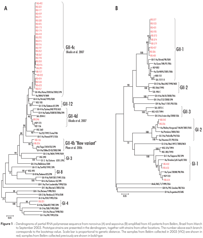
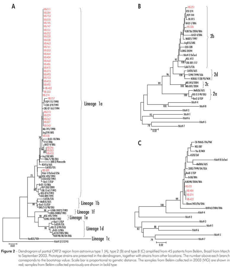
From a total of 305 samples tested by RT-PCR, 96 (31.5%) showed positive results, with 51 classified as HuCVs, 40 as HAstVs and five as mixed infections. Of the 56 (18.4%) samples that were positive for HuCV, 45 were sequenced: 30 (66.7%) were classified as NoVs in genogroups GI-4 (1, 3.3%) and GII-4 (29, 96.7%). Another 15 (33.3%) were identified as SaVs of types GI-1 (3, 20%), GI-2 (2, 13.3%) and GII-1 (10, 66.7%) (Figure 1, Table 1). The 11 samples that showed a faint band (low amplicon concentration) in the PCR were retested using NoV-specific primers; eight of the samples yielded a specific amplicon and three were negative. After sequencing, one of these samples showed positive results for GII-4. In the other seven samples, some nucleotides could not be defined and these strains were therefore not included in the dendrogram. Of the 45 (14.7%) samples classified as HAstVs, 37 (82.2%) belong to genotype 1, 4 (8.9%) to genotype 8, 2 (4.4%) to genotype 2, and two (4.4%) were not sequenced (Figure 2, Table 1). Five mixed infections were observed; four involved NoV/GII-4 plus HAstV-1 and one involved SaV/GII-1 plus HAstV-8.


The strains classified as NoVs GII-4 were grouped in three distinct sub-lineages: GII-4b/"new variant" (2/29; 6.9%), GII-4c (26/29; 89.6%) and GII-4d (1/29; 3.4%). The mean divergence observed among the strains classified as NoVs GII-4c and GII-4b/"new variant" was 0.5% and 2.9%, respectively. The divergences from the specific prototype were 3.1% (GII-4b "new variant"), 3% (GII-4c), 4.9% (GII-4d) and 11.3% (GI-4). The analysis involving only the strains from Belém demonstrated a mean divergence of 9.6% between GII-4c and GII-4d, and 16.4% between GII-4b and GII-4d. In relation to GI-4, this variation was higher than 49.3%. Little divergence was observed when the strains classified as genogroup GII-4 were analyzed in relation to the amino acid sequence (data not shown).
Among the SaVs, there was slight variation within each of the three genogroups detected: GII-1 (0.9%), GI-2 (0.3%) and GI-1 (3.1%). In relation to the prototypes the mean divergence was: GII-1 (4.1%), GI-2 (10.3%) and GI-1 (10.3%). Using the amino acid sequence of the Bristol prototype (AJ 249939; amino acid 1460 to 1555) as a reference, many substitutions were noted, characterizing the several genogroups / genotypes analyzed. However, no exclusive substitution was observed with respect to the strains from Belém (data not shown).
All of the HAstV strains detected in this study belonged to the lineage 1a and diverged from 0%-3.6% (mean of 0.6%) and 2.7% from the prototype HAstV-1 Oxford. No significant amino acid substitution was observed for the analyzed region (data not shown).
The samples tested in this study came from 21 hospitals and 11 emergency departments. A comparison of the positive rates obtained in the hospitals for NoVs (13.2% - 37/281), SaVs (5.3% - 15/281) and HAstVs (14.6% - 41/281) with those obtained in the emergency departments - 4.2% (1/24), 0% and 16.7% (4/24), respectively - revealed no statistically significant difference when using the Fisher exact test (p< 0.05). However, If NoVs and SaVs were taken together, (like HuCVs), the difference was significant (p = 0.04).
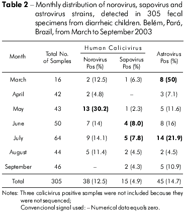
Table 2 showed the percentage of positives for NoVs, SaVs and HAstVs in gastroenteritis cases observed throughout the study period (March to September 2003). Peak incidence rates were verified: NoVs in May (13/43; 30.2%); SaVs in June (4/50; 8.0%) and July (5/64; 7.8%); and HAstVs in March (8/16; 50%) and July (14/64; 21.9%).

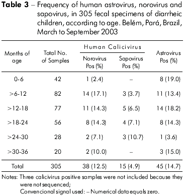
The age distribution of the NoV, SaV and HAstV-positive cases are shown in Table 3. The highest detection rate of NoVs was observed in children aged 6 to 12 months (17.1%); for SaVs this age was higher, 24 to 30 months-old (10.7%). Rates of HAstVs were comparable (19.0% and 18.2%) in 0- to 6- and 12- to 18-month-olds.

Most of the specimens were collected within 42 h of either hospitalization or health unit attendance. The main symptoms observed in the positive cases for these viruses were diarrhea, vomiting, fever and dehydration, but similar symptoms were found in negative cases (data not shown).
DISCUSSION
In the current study, at least one viral agent was detected in 31.5% of the stool samples tested. This result is slightly higher than 29% obtained in a five-year survey conducted in Gyeonggi Province, South Korea22. The detection rates obtained in Belém for HuCVs, NoVs, SaVs and HAstVs antigens were 18.4%, 12.5%, 4.9% and 14.7%, respectively. Studies conducted in hospitals in Campo Grande (7.6%), Goiânia and Brasília-DF (8.6%) and Salvador (9%) yielded lower HuCV detection rates than those obtained in Belém1,4,48. For NoVs, the rate observed in our study (12.5%) was lower than these registered in children from Korea (36.2%) and Espírito Santo (39.7%), but similar to those found in two studies conducted in Rio de Janeiro (14.5% and 20%) and São Paulo (15.7%) and higher than those of a study conducted in Thailand (6.5%)24,26,28,37,41,44.
Research on SaVs in Brazil is still limited because and most of the studies used primers that are specific to NoVs. However, these viruses were detected in one sample in Salvador (0.7%) and already described in diarrheic samples collected in a hospital (5.9%) and in a health unit (3%) in Belém at percentages similar to the ones presented in this work (4.9%)29,40,48. The rates of SaVs were similar to those reported in Thailand and Australia (4.8% and 4.1%, respectively) and lower than rates in India (10.2%)20,21,35. Three samples that were positive for HuCVs in our study gave negative results with specific primers for NoVs. This suggests that they probably belong to the genus SaVs, increasing the detection of these viruses to 5.9%.
The overall HAstV positive rate (14.7%) was similar to those recorded from outpatients and/or inpatients in South Korea, Cordoba City and Rio de Janeiro (11.9%, 12.4% and 14%, respectively)19,22,45. This percentage was higher than rates obtained previously in different studies conducted in Belém over 18 years, involving hospitals, health units, community and a day care center, which varied from 2.7% to 9.9%15.
The NoVs dendrogram shown in this study was constructed based on the sequences obtained with primers 289/290, which encode part of the RNA polymerase. These two primers were used because they detect both NoVs and SaVs. In contrast, most other studies conducted in Brazil used other pairs of primers that amplify region B of the RNA polymerase and are specific to NoVs. Therefore, we compared the study carried out in Belém with others from Brazil, as these primers amplified the same RNA polymerase region. In our study, GII-4 was detected in 96.7% of the strains analyzed. These results are consistent with those obtained in Rio de Janeiro (64%) and in São Paulo (76.9%)7,44. GII-4 was also the prevalent genotype that circulated in Belém in another study of samples collected in the years of 1992-1994 and 1998-2000 (data not shown). Many articles report a marked increase on the circulation of GII-4 strains since the 1990s, with growing genetic variability, that were related either with extensive outbreaks and sporadic cases5,31,43.
Three different genotypes of SaVs circulated during this study; GII-1 was the most prevalent (66.7%). This genotype was also detected in one sample from Salvador, the only place in Brazil where this virus was also found48. In contrast, GI-1 was the most prevalent strain in previous studies conducted in Belém40. A similar occurrence of GI-1 was also observed in Bangladesh among diarrheic children8.
Three HAstVs genotypes co-circulated in Belém during the six months of study with HAstV-1 as the most prevalent genotype (82.2%), as reported elsewhere6,16,39,45. The concurrent circulation of other genotypes was observed in various settings in Brazil as well as across the globe6,10,36,39. All HAstV-1 positive samples were classified as lineage 1a. Previous studies conducted in Belém demonstrated that 93.8% (76/81) of HAstV-1 strains detected over a period of 18 years (1982-2000) were classified as 1a15. However, studies conducted in other regions of Brazil, such as Rio de Janeiro and Goiânia, from 2003 to 2005, demonstrated the circulation of lineage 1 d38,45. The data obtained in this study, from samples collected in 2003, showed that no change had occurred in Belém, as the 1a lineage continue to be the only in circulation. Interestingly, HAstV-8 was detected in 8.9% of the samples. This genotype is considered rare though it had already been observed in other studies conducted in Belém with less frequency and similar results were observed in Rio de Janeiro17,45.
Notably, co-infection involving the three viruses was observed in 1.6% of cases. These situations are relatively common in gastroenteritis episodes, as described by other authors9,18,25,36,44. This fact probably reflects the poor sanitation conditions and the low socioeconomic environment in which these children live.
This study involved samples collected either in hospitals or in emergency departments. A higher detection frequency for these viruses was observed in the hospitals, indicating a greater severity of these agents associated with gastroenteritis. However, the number of specimens (42) collected in the emergency departments was too small to allow better analyzes.
We also observed that NoVs were more prevalent than SaVs in children from the ages of 6 to 12 months, while SaVs were more prevalent in children and from 24 to 30 months old. These data are consistent with the literature, which indicates that SaVs are more frequently detected in older children12. A seasonality pattern for these three agents could not be defined as the study involved only six months of research.
In conclusion, we demonstrated the relevance of these viruses to acute gastroenteritis in hospitals and in emergency departments. The detection of SaVs reinforces the need to establish it monitoring systems to evaluate the impact of this virus in other regions of Brazil. The molecular characterization of these agents demonstrated that different genotypes circulate over time (i.e., were not the same in this study and in previous studies conducted in Belém). Additional molecular studies are therefore necessary to monitor these variations. The recent introduction of an RV-A vaccine in the Pediatric National Immunization Program in Brazil further highlights the need to continue research on these viruses.
ACKNOWLEDGMENTS
We gratefully acknowledge the valuable technical support provided by Maria Silvia Sousa de Lucena, Jones Anderson Monteiro Siqueira, Ian Carlos Lima, Dielle Monteiro Teixeira and Jefferson Oliveira. Thanks are also due to the field staff and doctors that worked during the project "Vigilância Intensiva das Diarréias em Hospitais". This work was supported by IEC/SVS/MS.
REFERENCES
1 Andreasi MS, Cardoso DD, Fernandes SM, Tozetti IA, Borges AM, Fiaccadori FS, et al. Adenovirus, calicivirus and astrovirus detection in fecal samples of hospitalized children with acute gastroenteritis from Campo Grande, MS, Brazil. Mem Inst Oswaldo Cruz. 2008 Nov;103(7):741-4. DOI:10.1590/S0074-02762008000700020 [ Links ]
2 Atmar RL, Estes MK. Diagnosis of noncultivatable gastroenteritis viruses, the human caliciviruses. Clin Microbiol Rev. 2001 Jan;14(1):15-37. DOI: 10.1128/CMR.14.1.15–37.2001 [ Links ]
3 Boom R, Sol CJ, Salimans MM, Jansen CL, Wertheim-van Dillen PM, Noordaa J. Rapid and simple method for purification of nucleic acids. J Clin Microbiol. 1990 Mar;28(3):495-503. [ Links ]
4 Borges AM, Teixeira JM, Costa PS, Giugliano LG, Fiaccadori FS, Franco RC, et al. Detection of calicivirus from fecal samples from children with acute gastroenteritis in the West Central region of Brazil. Mem Inst Oswaldo Cruz. 2006 Nov;101(7):721-4. DOI:10.1590/S0074-02762006000700003 [ Links ]
5 Bull RA, Tu ET, McIver CJ, Rawlinson WD, White PA. Emergence of a new norovirus genotype II. 4 variant associated with global outbreaks of gastroenteritis. J Clin Microbiol. 2006 Feb;44(2):327-33. DOI: 10.1128/JCM.44.2.327-333.2006 [ Links ]
6 Cardoso DDP, Fiaccadori FS, Souza MBLD, Martins RM, Leite JPG. Detection and genotyping of astroviruses from children with acute gastroenteritis from Goiania, Goiás, Brazil. Med Sci Monit. 2002 Sep;8(9):CR624-8. [ Links ]
7 Castilho JG, Munford V, Resque HR, Fagundes-Neto U, Vinjé J, Rácz ML. Genetic diversity of norovirus among children with gastroenteritis in São Paulo State, Brazil. J Clin Microbiol. 2006 Nov;44(11):3947-53. DOI:10.1128/JCM.00279-06 [ Links ]
8 Dey SK, Phan TG, Nguyen TA, Nishio O, Salim AF, Yagyu F, et al. Prevalence of sapovirus infection among infants and children with acute gastroenteritis in Dhaka City, Bangladesh during 2004-2005. J Med Virol. 2007 May;79(5):633-8. [ Links ]
9 Dove W, Cunliffe NA, Gondwe JS, Broadhead RL, Molyneux ME, Nakagomi O, et al. Detection and characterization of human caliciviruses in hospitalized children with acute gastroenteritis in Blantyre, Malawi. J Med Virol. 2005 Dec;77(4):522-7. [ Links ]
10 Espul A, Martinez N, Noel JS, Cuello H, Abrile C, Grucci S, et al. Prevalence and characterization of astroviruses in Argentinean children with acute gastroenteritis. J Med Virol. 2004 Jan;72(1):75-82. [ Links ]
11 Fankhauser RL, Monroe SS, Noel JS, Humphrey CD, Bresee JS, Parashar UD, et al. Epidemiological and molecular trends of "Norwalk-like viruses" associated with outbreaks of gastroenteritis in the United States. J Infect Dis. 2002 Jul;186(1):1-7. [ Links ]
12 Farkas T, Deng X, Ruiz-Palacios G, Morrow A, Jiang X. Development of an enzyme immunoassay for detection of sapovirus-specific antibodies and its application in a study of seroprevalence in children. J Clin Microbiol. 2006 Oct;44(10):3674-9. DOI:10.1128/JCM.01087-06 [ Links ]
13 Fauquet CM, Mayo MA, Maniloff J, Desselberger U, Ball LA. Family Astroviridae. In: Fauquet CM, Mayo MA, Maniloff J, Desselberger U, Ball LA, editors. Virus taxonomy: eighth report of the International Committee on the taxonomy of viruses. San Diego: Elsevier Academic Press; 2005. p. 859-64.
14 Felsenstein J. PHYLIP, Version 3.57c. Seatle, WA: Department of Genetics, University of Washington; 1995.
15 Gabbay YB, Leite JP, Oliveira DS, Nakamura LS, Nunes MR, Mascarenhas JD, et al. Molecular epidemiology of astrovirus type 1 in Belém, Brazil, as an agent of infantile gastroenteritis, over a period of 18 years (1982-2000): identification of two possible new lineages. Virus Res. 2007 Nov;129(2):166-74. [ Links ]
16 Gabbay YB, Linhares AC, Cavalcante-Pepino EL, Nakamura LS, Oliveira DS, Silva LD, et al. Prevalence of human astrovirus genotypes associated with acute gastroenteritis among children in Belém, Brazil. J Med Virol. 2007 May;79(5):530-8. [ Links ]
17 Gabbay YB, Linhares AC, Oliveira DS, Nakamura LS, Mascarenhas JD, Gusmão RH, et al. First detection of a human astrovirus type 8 in a child with diarrhea in Belém, Brazil: comparison with other strains worldwide and identification of possible three lineages. Mem Inst Oswaldo Cruz. 2007 Jun;102(4):531-4. DOI:10.1590/S0074-02762007005000032 [ Links ]
18 Gabbay YB, Luz CR, Costa IV, Cavalcante-Pepino EL, Sousa MS, Oliveira KK, et al. Prevalence and genetic diversity of astroviruses in children with and without diarrhea in São Luís, Maranhão, Brazil. Mem Inst Oswaldo Cruz. 2005 Nov;100(7):709-14. DOI: 10.1590/S0074-02762005000700004 [ Links ]
19 Giordano MO, Martinez LC, Isa MB, Paez Rearte M, Nates SV. Childhood astrovírus-associated diarrhea in the ambulatory setting in a public hospital in Cordoba City, Argentina. Rev Inst Med Trop Sao Paulo. 2004 Mar-Apr;46(2):93-6. DOI: 10.1590/S0036-46652004000200007 [ Links ]
20 Hansman GS, Katayama K, Maneekarn N, Peerakome S, Khamrin P, Tonusin S, et al. Genetic diversity of norovirus and sapovirus in hospitalized infants with sporadic cases of acute gastroenteritis in Chiang Mai, Thailand. J Clin Microbiol. 2004 Mar;42(3):1305-7. DOI: 10.1128/JCM.42.3.1305-1307.2004 [ Links ]
21 Hansman GS, Takeda N, Katayama K, Tu ET, Mclver CJ, Rawlinson WD, et al. Genetic diversity of sapovirus in children, Australia. Emerg Infect Dis. 2006 Jan;12(1):141-3. [ Links ]
22 Huh JW, Kim HW, Moon SG, Lee JB, Lim YH. Viral etiology and incidence associeted with acute gastroenteritis in a 5-year survey in Gyeonggi province, South Korea. J Clin Virol. 2009 Feb;44(2):152-6. [ Links ]
23 Jiang X, Huang PW, Zhong WM, Farkas T, Cubitt DW, Matson DO. Design and evaluation of a primer pair that detects both Norwalk- and Sapporo-like caliciviruses by RT-PCR. J Virol Methods. 1999 Dec;83(1-2):145-54. [ Links ]
24 Kittigul L, Pombubpa K, Taweekate Y, Yeephoo T, Khamrin P, Ushijima H. Molecular characterization of rotaviruses, noroviruses, sapovirus, and adenoviruses in patients with acute gastroenteritis in Thailand. J Med Virol. 2009 Feb;81(2):345-53. [ Links ]
25 Klein EJ, Boster DR, Stapp JR, Wells JG, Qin X, Clausen CR, et al. Diarrhea etiology in a Children's Hospital Emergency Department: a prospective cohort study. Clin Infect Dis. 2006 Oct;43(7):807-13. DOI: 10.1086/507335 [ Links ]
26 Koh H, Baek SY, Shin JI, Chung KS, Je YM. Coinfection of viral agents in Korean children with acute watery diarrhea. J Korean Med Sci. 2008 Dec;23(6):937-40.
27 Méndez E, Arias CF. Astroviruses. In: Knipe DM, Howley PM, editors. Fields virology. 5th ed. Philadelphia: Lippincott Williams and Wilkins; 2007. p. 981-1000.
28 Morillo SG, Cilli A, Carmona RC, Timenetsky MC. Identification and molecular characterization of norovirus in São Paulo State, Brazil. Braz J Microbiol. 2008 Dec;39(4):619-22. DOI:10.1590/S1517-83822008000400004 [ Links ]
29 Nakamura LS, Oliveira DS, Silva PF, Lucena MS, Mascarenhas JD, Gusmão RH, et al. Molecular characterization of calicivirus in feces of children with acute diarrhea, attending a public hospital, in Belém, Pará. In: XVII National Meeting of Virology; 2006 19-22; Campos do Jordão: Sociedade Brasileira de Virologia; 2006. p. 95. (Virus Reviews & Research; vol. 11; supl. 1).
30 Noel JS, Lee TW, Kurtz JB, Glass RI, Monroe SS. Typing of human astroviruses from clinical isolates by enzyme immunoassay and nucleotide sequencing. J Clin Microbiol. 1995 Apr;33(4):797-801. [ Links ]
31 Okada M, Ogawa T, Yoshizumi H, Kubonoya H, Shinozaki K. Genetic variation of the norovirus GII-4 genotype associated with a large number of outbreaks in Chiba prefecture, Japan. Arch Virol. 2007;152(12):2249-52. DOI: 10.1007/s00705-007-1028-8 [ Links ]
32 Parashar UD, Gibson CJ, Breese JS, Glass RI. Rotavirus and severe childhood diarrhea. Emerg Infect Dis. 2006 Feb;12(2):304-6. [ Links ]
33 Patel MM, Widdowson MA, Glass RI, Akazawa K, Vinjé J, Parashar UD. Systematic literature review of role of noroviruses in sporadic gastroenteritis. Emerg Infect Dis. 2008 Aug;14(8):1224-31. DOI: 10.3201/eid1408.071114 [ Links ]
34 Phan TG, Khamrin P, Quang TD, Dey SK, Takanashi S, Okitsu S, et al. Emergence of intragenotype recombinant sapovirus in Japan. Infect Genet Evol. 2007 Jul;7(4):42-6. DOI: 10.1016/j.meegid.2007.02.004 [ Links ]
35 Rachakonda G, Choudekar A, Parveen S, Bhatnagar S, Patwari A, Broor S. Genetic diversity of noroviruses and sapoviruses in children with acute sporadic gastroenteritis in New Delhi, India. J Clin Virol. 2008 Sep;43(1):42-8. DOI:10.1016/j.jcv.2008.05.006 [ Links ]
36 Resque HR, Munford V, Castilho JG, Schmich H, Caruzo TA, Rácz ML. Molecular characterization of astrovirus in stool samples from children in São Paulo, Brazil. Mem Inst Oswaldo Cruz. 2007 Dec;102(8):969-74. DOI: 10.1590/S0074-02762007000800012 [ Links ]
37 Ribeiro LR, Giuberti RS, Barreira DM, Saick KW, Leite JP, Miagostovich MP, et al. Hospitalization due to norovirus and genotypes of rotavirus inpediatric patients, state of Espírito Santo. Mem Inst Oswaldo Cruz. 2008 Mar;103(2):201-6. DOI: 10.1590/S0074-02762008000200013 [ Links ]
38 Silva PA, Cardoso DD, Schreier E. Molecular characterization of human astroviruses isolated in Brazil, including the complete sequences of astrovirus genotypes 4 and 5. Arch Virol. 2006 Jul;151(7):1405-17. DOI 10.1007/s00705-005-0704-9 [ Links ]
39 Silva PA, Santos RA, Costa PS, Teixeira JM, Giugliano LG, Andreasi MS, et al. The circulation of human astrovirus genotypes in the Central West Region of Brazil. Mem Inst Oswaldo Cruz. 2009 Jul;104(4):655-8. DOI: 10.1590/S0074-02762009000400021 [ Links ]
40 Siqueira JM, Aragão GC, Nascimento IS, Oliveira DS, Santos MC, Lima IC, et al. Estudos da prevalência de calicivírus humanos em crianças com gastroenterite atendidas em um posto de saúde de Belém-Pará no período de 1998-1999. In: Anais do II Congresso Norte Nordeste de Infectologia; 2008 nov. 28-30; Belém: Sociedade Brasileira de Infectologia; 2008.
41 Soares CC, Santos N, Beard RS, Albuquerque MC, Maranhão AG, Rocha LN, et al. Norovirus Detection and genotyping for children with gastroenteritis, Brazil. Emerg Infect Dis. 2007 Aug;13(8):1244-6. [ Links ]
42 Tamura K, Dudley J, Nei M, Kumar S. MEGA4: Molecular Evoutinary Genetics Analysis (MEGA) software version 4.0. Mol Biol Evol. 2007 Aug;24(8):1596-9. DOI:10.1093/molbev/msm092 [ Links ]
43 Tu ET, Bull RA, Greening GE, Hewitt J, Lyon MJ, Marshall JA, et al. Epidemics of gastroenteritis during 2006 Were Associated with the Spread of Norovirus GII.4 Variants 2006a and 2006b. Clin Infect Dis. 2008 Feb;46(3):413-20. DOI: 10.1086/525259 [ Links ]
44 Victoria M, Carvalho-Costa FA, Heinemann MB, Leite JP, Miagostovich M. Prevalence and molecular epidemiology of noroviruses in hospitalized children with acute gastroenteritis in Rio de Janeiro, Brazil, 2004. Pediatr Infect Dis J. 2007 Jul;26(7):602-6. [ Links ]
45 Victoria M, Carvalho-Costa FA, Heinemann MB, Leite JP, Miagostovich MP. Genotypes and molecular epidemiology of human astroviruses in hospitalized children with acute gastroenteritis in Rio de Janeiro, Brazil. J Med Virol. 2007 Jul;79(7):939-44. [ Links ]
46 Walter JE, Mitchell DK, Guerrero ML, Berke T, Matson DO, Monroe SS, et al. Molecular epidemiology of human astrovirus diarrhea among children from a Periurban community of Mexico City. J Infect Dis. 2001 Mar;183(5):681-6. [ Links ]
47 Wilhelmi I, Roman E, Sánchez-Fauquier A. Viruses causing gastroenteritis. Clin Microbiol Infect. 2003 Apr;9(4):247-62. [ Links ]
48 Xavier MP, Oliveira SA, Ferreira MS, Victoria M, Miranda V, Silva MF, et al. Detection of caliciviruses associated with acute infantile gastroenteritis in Salvador, an urban center in Northeast Brazil. Braz J Med Biol Res. 2009 May;42(5):438-44. DOI: 10.1590/S0100-879X2009000500007 [ Links ]
 Corresponding / Correspondência / Correspondencia:
Yvone Benchimol Gabbay
Instituto Evandro Chagas, Seção de Virologia
Rodovia BR316, km 7, s/nº, Levilândia
CEP: 67.030-000 Ananindeua, Pará, Brasi
E-mail:yvonegabbay@iec.pa.gov.br
Recebido em / Received / Recibido en: 31/7/2009
Aceito em / Accepted / Aceito en: 19/10/2009


 texto em
 Curriculum ScienTI
