


Serviços Personalizados
Journal

Artigo
 Inglês (pdf)
 Artigo em XML
 Referências do artigo
 Enviar este artigo por email
Indicadores
-
 Citado por SciELO
Links relacionados
-
 Similares em
SciELO
Similares em
SciELO
Compartilhar

 Permalink
Revista Pan-Amazônica de Saúde
versão impressa ISSN 2176-6215versão On-line ISSN 2176-6223
Rev Pan-Amaz Saude v.3 n.1 Ananindeua mar. 2012
http://dx.doi.org/10.5123/S2176-62232012000100008
CASE REPORT | RELATO DE CASO | RELATO DE CASO
Anetoderma secondary to borderline leprosy
Anetodermia secundária à hanseníase dimorfa
Anetodermia secundaria a hanseniasis dimorfa
Taurino dos Santos Rodrigues NetoI; Francisca Regina de Oliveira CarneiroII; Caren dos Santos LimaI; Heliana Freitas de Oliveira GóesI; Natália Borges NunesI
IUniversidade do Estado do Pará, Belém, Pará, Brasil
IIAmbulatório de Dermatologia, Universidade do Estado do Pará, Belém, Pará, Brasil
Endereço para correspondência
Correspondence
Dirección para correspondencia
ABSTRACT
OBJECTIVE: To report a case of secondary anetoderma in a patient with borderline leprosy and thus draw attention to this unusual association of anetoderma and hanseniasis.
CASE REPORT: A mixed-race man who was 21 years old was referred to the Ambulatory of Dermatology of Universidade do Estado do Pará, in the City of Belém, Pará State, Brazil, with a diagnosis of borderline leprosy at 10 years old. Years later, the patient developed "scrolling" surface scars, with a diagnosis compatible with anetoderma lesions secondary to Hansen's disease.
FINAL CONSIDERATIONS: A dermatological affection, although rare, may be associated with common systemic diseases, such as leprosy, and the disclosure of such affections is emphasized in this case. Their investigation and exclusion are necessary when faced with such conditions.
Keywords: Anetoderma; Leprosy; Skin Diseases, Bacterial; Elastic Tissue.
RESUMO
OBJETIVO: Relatar um caso de anetodermia secundária em um paciente com hanseníase dimorfa e, consequentemente, chamar atenção para esta associação incomum entre anetodermia e hanseníase.
RELATO DE CASO: Um homem de cor parda, com 21 anos de idade, foi encaminhado para o Ambulatório de Dermatologia da Universidade do Estado do Pará, na Cidade de Belém, Estado do Pará, Brasil, com um diagnóstico de hanseníase dimorfa desde os 10 anos de idade. Depois de alguns anos, o paciente desenvolveu cicatrizes de superfície apergaminhada com diagnóstico compatível com lesões decorrentes de anetodermia secundária à hanseníase.
CONSIDERAÇÕES FINAIS: Uma afecção dermatológica, embora rara, pode ser associada a doenças sistêmicas comuns, como a hanseníase, e a descoberta dessas afecções é então enfatizada. Sua investigação e exclusão são necessárias quando confrontadas com tais circunstâncias.
Palavras-chave: Anetodermia; Hanseníase; Dermatopatias Bacterianas; Tecido Elástico.
RESUMEN
OBJETIVO: Relatar un caso de anetodermia secundaria en un paciente con hanseniasis dimorfa y, consecuentemente, llamar la atención sobre asociación no común entre anetodermia y hanseniasis.
RELATO DE CASO: un hombre pardo, de 21 años de edad, fue encaminado al Ambulatorio de Dermatología de la Universidad del Estado de Pará, en la ciudad de Belém, Estado de Pará, Brasil, con un diagnóstico de hanseniasis dimorfa desde los 10 años de edad. Después de algunos años, el paciente desarrolló cicatrices de superficie apergaminada con diagnóstico compatible con lesiones decurrentes de anetodermia secundaria a la hanseniasis.
CONSIDERACIONES FINALES: una afección dermatológica, aunque rara, puede ser asociada a enfermedades sistémicas comunes, como la hanseniasis, enfatizándose el hallazgo de dichas afecciones. La investigación y la exclusión de la misma se hacen necesarias cuando confrontadas con tales circunstancias.
Palabras clave: Anetodermia; Lepra; Enfermedades Cutáneas Bacterianas; Tejido Elástico.
INTRODUCTION
Anetodermas (from the Greek: anetos = flat; derma = skin) were described in 1882 by Jadassohn, who used the word to describe the clinical-pathological feature of a new disease in a patient with erythematous maculae that had evolved into scars1,2,3. They have an unknown etiology4, but possible causes include auto-immune mechanisms5 (lupus and scleroderma), genetics causes6 (familial forms), infectious processes1 (chronic atrophying acrodermatitis associated with borreliosis, hanseniasis, syphilis, varicella, and tuberculosis), defects in the synthesis of elastic fibers7 (penicillamine inhibiting lysiloxydase) and elastophagocytosis4. We report the case of a patient who presented anetoderma lesions secondary to borderline leprosy (BL), an association that is considered rare.
CASE REPORT
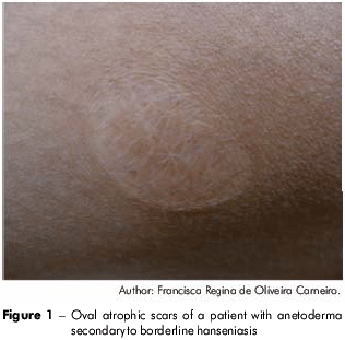
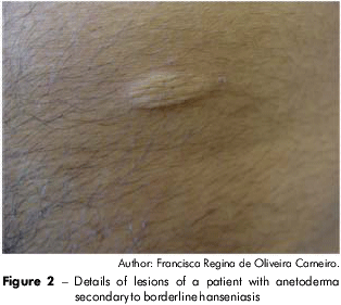
A male, 21 years old, originally from Belém, Pará State, Brazil, presented atrophic and circular lesions for two years distributed along the trunk and arms, with "scrolling" surface scars that varied in size from 3 to 4 cm. The lesions were asymptomatic and had been growing in number and size (Figures 1 and 2).
This project was approved by the Universidade do Estado do Pará (UEPA) Centro de Ciências Biológicas e da Saúde (CCBS) human research ethics committee (01330352000-09/November-2009).
Skin smears were performed for the lesions and were negative for acid-fast bacilli. A biopsy and histopathological exam revealed epidermis with intergemmal crests and dermis presenting a discreet lymphocytic infiltrate around the bases of the superior segment and compact hyaline of the conjunctiva at the inferior segment aspect. Verhoff staining showed the absence of elastic fibers in all superior dermis, which was compatible with anetoderma.
VDRL and serologies for HIV, rubella and hepatitis were all negative. Anti-nuclear factor (AFN) and anti-native DNA antibodies were not reactive.
The patient also reported that at 10 years old, he was diagnosed with borderline leprosy and had received 24 doses of multi-bacillary multidrug therapy (MDT/WHO); he was discharged as cured. However, he denied a personal history of tuberculosis, chickenpox or other infectious diseases.
DISCUSSION
Anetodermas, also known as macular atrophies, are characterized by atrophic lesions, which are generally oval or circular, due to the destruction of the elastic dermal tissue. They may be classified into three groups: hereditary, primary and secondary8.
They affect men and women equally, and the average age of appearance is 20 years. Clinically, they vary from 1 cm to more than 10 cm, and their number varies from a few to hundreds. They are recognized as areas of flat tissue covered with thin, hypochromic and herniated skin that may be protruded due to their flatness8. They are located in the trunk, arms, and nape and are able to affect the face. Their etiology is unknown, but they are associated with an increase in elastolysis. Moreover, they have a chronic evolution and a high resistance to treatment2,9.
Anetodermas may be classified as idiopathic Schwenninger-Buzzi anetoderma, idiopathic Jadassohn anetoderma, Pellizari anetoderma, hereditary macular atrophy, which evolves into anetoderma scars with brilliant hypochromic and superficial atrophy6, and secondary anetoderma, which is more frequent2.
Secondary anetoderma occurs in patients with anterior cutaneous inflammatory processes, such as lupus, syphilis, Hansen's disease, tuberculosis, varicella, and borreliosis2. In this case, the lesions do not necessarily appear at the same place as the cutaneous manifestation of the associated disease.
The diagnosis is essentially clinical and based on anamnesis. It is confirmed by a histopathological exam revealing superficial and profound lymphocytic or vascular infiltrate, with a predominance of lymphocytes. Epidermal alterations are not observed, not even in the subcutaneous cellular tissue7. Granulomas may be found in giant cells in the dermis7,10. Diagnostic confirmation requires special staining methods to identify the elastic fibers (orcein, Verhoff-Van Gieson), elastolysis and elastorrhexis, which affect the papillar and reticular dermis. The remaining fibers are fragmented, adopting a characteristic tortuous and thin aspect7.
Achenbach et al9 described a case of anetoderma secondary to polar tuberculoid hanseniasis, and they observed a hypochromic area of 7x5 cm located on the anterior face of the left arm that was protruded, with a hernia and thin skin, which corresponded to the first plaque of polar tuberculoid hanseniasis. When this plaque became involuted after receiving proper treatment, the lesion became anetodermic.
Therefore, despite the rarity of this association, the present case illustrates the aspects of this affection, highlighting the importance of further studies to determine the exact correlations between cytokines and antigens in such associations.
FINAL CONSIDERATIONS
This case emphasizes the clinical presentation of a rare dermatological condition and the importance of its recognition and research to exclude the diagnosis of systemic diseases that are sometimes associated.
REFERENCES
1 Karrer S, Szeinies RM, Stolz W, Landthaler M. Primary anetoderma in children: report of two cases and literature review. Pediatr Dermatol. 1996;13(5):382-5. Doi:10.1111/j.1525-1470.1996.tb00705.x [Link]
2 Coelho WS, Gurtler TGR, Diniz IM, Souza Filho JB. Caso para diagnóstico. An Bras Dermatol. 2008;83(6):578-80. http://dx.doi.org/10.1590/S0365-05962008000600015 [Link]
3 Chargin L, Silver H. Macular Atrophy of the skin. Arch Dermat Syph. 1931;24:614-43. doi:10.1001/archderm.1931.01450010624012 [Link]
4 Zaki I, Scerri L, Nelson H. Primary anetoderma: phagocytosis of elastic fibers by macrophages. Clin Exp Dermatol. 1994;19(5):388-90. [Link]
5 Hodak E, Shamai-Lubotitz O, David M, Hazaz B, Lahav M, Sandbank M. Primary anetoderma with a wide spectrum of autoimmune abnormalities. J Am Acad Dermatol. 1991;25(2 Pt 2):415-8.
6 Friedman SJ, Venencie PY, Bradley RR, Winkelmann RK. Familial anetoderma. J Am Acad Dermatol. 1987;16(2 Pt 1):341-5.
7 Venencie PY, Winkelmann RK, Moore BA. Anetoderma. Arch Dermatol. 1984;120:1032-8.
8 Azulay RD, Azulay DR, Abulafia LA. Dermatologia. 5. ed. Rio de Janeiro: Guanabara Koogan; 2008.
9 Achenbach R, Jorge M, Schroh RG, Liturri M, Corbella MC. Anetodermia secundaria a una lepra tuberculoide polar. Arch Argent Dermatol. 2002;52(1):27-30.
10 Ishida Y, Naitoh M, Yoneyama K, Shinkai H, Miyachi Y, Utani A. Coexistence of disseminated primary anetoderma and generalized granuloma annulare-like papules. J Dermatol. 2007;34(4):278-9.
 Correspondence / Correspondência / Correspondencia:
Taurino dos Santos Rodrigues Neto
Rua Boaventura da Silva, 1227, apt. 1302.
Bairro: Umarizal
CEP: 66060-060
Belém-Pará-Brasil
Tel.: +55 (91) 3241-8430
E-mail: taurinorodrigues@gmail.com
Received / Recebido em /Recibido en: 4/6/2012
Accepted / Aceito em / Aceito en: 27/11/2012
