


Servicios Personalizados
Revista

Articulo
 texto en
  Inglés (pdf)
 Articulo en XML
 Referencias del artículo
 Curriculum ScienTI
 Enviar articulo por email
Indicadores
-
 Citado por SciELO
Links relacionados
-
 Similares en
SciELO
Similares en
SciELO
Compartir

 Permalink
Revista Pan-Amazônica de Saúde
versión impresa ISSN 2176-6215versión On-line ISSN 2176-6223
Rev Pan-Amaz Saude vol.12 Ananindeua 2021 Epub 26-Jul-2021
http://dx.doi.org/10.5123/s2176-6223202100820
CASE REPORT
Late diagnosis of mycosis fungoides: a case report
1 Universidade Federal do Pará, Núcleo de Medicina Tropical, Programa de Pós-Graduação em Doenças Tropicais, Belém, Pará, Brasil
2 Universidade do Estado do Pará, Centro de Ciências Biológicas e da Saúde, Laboratório de Pesquisas em Dermatoses de Interesse Sanitário, Belém, Pará, Brasil
3 Hospital Ophir Loyola, Divisão de Hematologia-Oncologia e Transplante de Células-Tronco, Belém, Pará, Brasil
4 Universidade Federal do Pará, Núcleo de Medicina Tropical, Laboratório de Pesquisas em Dermatologia Tropical e Doenças Endêmicas, Belém, Pará, Brasil
INTRODUCTION:
Mycosis fungoides (MF) is a cutaneous T-cell lymphoma with an incidence of approximately 0.41/100,000 individuals annually. This report highlights the impacts of delayed diagnosis on the evolution of the disease.
CASE REPORT:
A female patient sought medical assistance at the dermatology service of the Universidade do Estado do Pará (UEPA), Brazil, on November 2017, due to three infiltrated erythematous-hyperchromic plaques, two on the abdomen and one scaly on the left lower limb, and a palpable lymph node in the left inguinal region with progressive growth which started in 2011. The patient reported that she had undergone several tests and treatments and was seen by six medical doctors of different specialties and underwent four histopathological exams. The patient's case was investigated in the dermatology service of UEPA; an immunohistochemical study was performed, concluding the diagnosis of MF. She was treated with methotrexate 15 mg and folic acid 5 mg in a single weekly dose and phototherapy with PUVA, showing significant improvement after treatment, but with an extensive area of atrophy and secondary infections.
CONCLUSION:
The present case is a public health issue that reinforces the need for adequate medical training for early identification and treatment of MF.
Keywords: Mycosis Fungoides; Health Impact Assessment; Delayed Diagnosis; Health Services Accessibility
INTRODUCTION
Mycosis fungoides (MF) and Sézary syndrome are the most common types of cutaneous T-cell lymphoma (CTCL), representing 44 to 54% of cases. MF mainly affects adults, with an average age between 55 and 60 years old, but it can also occur in children and adolescents. It occurs in the proportion of 1.6-2.0 men to 1.0 women1,2.
The cause of MF is still not fully understood, but evidence suggests that it occurs due to chronic antigenic stimulation that can generate proliferation and accumulation of T cells in the skin. This stimulus is greatly influenced by the skin microenvironment, including dendritic cells and reactive cytotoxic or regulatory T cells3. The T lymphocytes involved in the etiopathogenesis of MF are CD4+ T cells that present adhesion molecules, such as CCR4 and CLA. Antigens such as CD7 or eventually CD5 or CD2 are reduced or absent in pathogenic cells4.
Patients with MF present, at first, mild skin lesions that may resemble eczema or diffuse erythema, which may progress to the tumor stage when there is an increase of lesion infiltration and, usually, ulceration. These lesions often occur in areas not frequently exposed to the sun, such as the torso, and may manifest with hypochromia or hyperchromia5. It is noteworthy that the diagnosis of this pathology is confirmed considering both clinical and histopathological criteria6.
The treatment is chosen according to the stage of the disease. In the initial stage (IA to IIA), skin-directed therapies (SDT) are used, such as topical corticosteroids, radiotherapy, and phototherapy, and in the advanced stage (IIB up to IVB), systemic therapy with retinoids, interferons, or immunobiologicals with SDT is applied7,8.
CASE DESCRIPTION
A 56-year-old female patient, housewife, born and raised in Belém, Pará State, Brazil, went to the dermatology service of the State University of Pará (Universidade do Estado do Pará - UEPA) in November 2017 due to a dark spot of progressive growth in the left lower limb (LLL), which appeared in May 2011. The patient also reported "swelling" and local pain episodes, besides two new spots on the abdomen and a swollen lymph node in the left inguinal region. She reported that she had been submitted to several tests and treatments without any improvement or diagnostic conclusion. In this context, the patient was seen by three general practitioners, an oncologist, and two dermatologists, with the first medical appointment a month after the beginning of symptoms but without a diagnostic conclusion. She was referred to the dermatology service on November 16, 2017 and got the final diagnosis on December 18, 2017, after six years and seven months of disease progression. Skin examination revealed two erythematous-hyperchromic infiltrated plaques in the abdomen, a desquamative erythematous-hyperchromic plaque with infiltration in the LLL, and a palpable lymph node in the left inguinal region under erythematous skin (Figures 1 and 2). Since the beginning of her condition, four histopathological examinations and one immunohistochemical examination of the lesion were performed, showing, finally, an atypical lymphoid infiltrate with epidermotropism, predominant CD4 T-cell immunophenotype, and loss of CD7 expression suggesting the diagnosis of MF. Computed tomography scans of the chest and abdomen showed no significant changes, and concomitant follow-up with hematology was performed. In addition to the palpable lymph node, swollen lymph nodes were found in the right inguinal region, after soft tissue ultrasound, with multiple lymph nodes involved, the largest one was 3.2 x 1 cm, in addition to skin thickening and fat densification. After evaluation, the staging was established as T2NxM0B0. Thus, the proposed treatment was methotrexate 15 mg and folic acid 5 mg, in a single weekly dose, and phototherapy with PUVA that started on 02/22/2018. The patient showed significant improvement in the lesions during the treatment, infiltration, erythema, desquamation, and ganglia have decreased, but with an extensive area of atrophy (Figure 3) and secondary infections with two episodes of erysipelas and one of impetigo in the affected limb, with intervals of four to six months between them. The erysipelas was treated with cephalexin 500 mg orally every 6 h for 14 days, and impetigo was treated with topical fusidic acid twice daily, improving the treatment after the administration of antibiotic therapy.
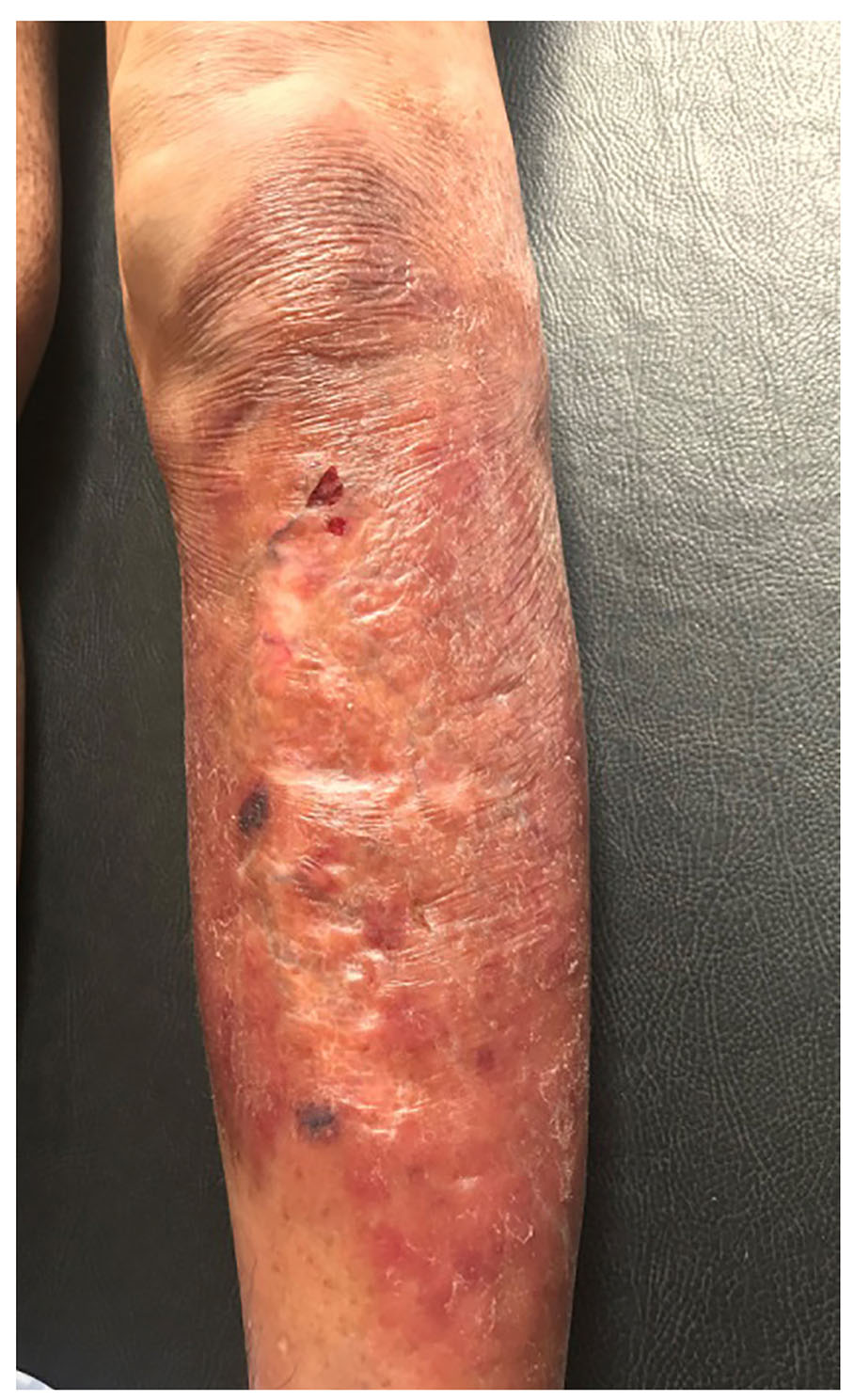
Photo: Brena Andrade de Lima Lobato.
Figure 1 - Infiltrated erythematous-hyperchromic plaque with desquamation and ulcers in the left lower limb
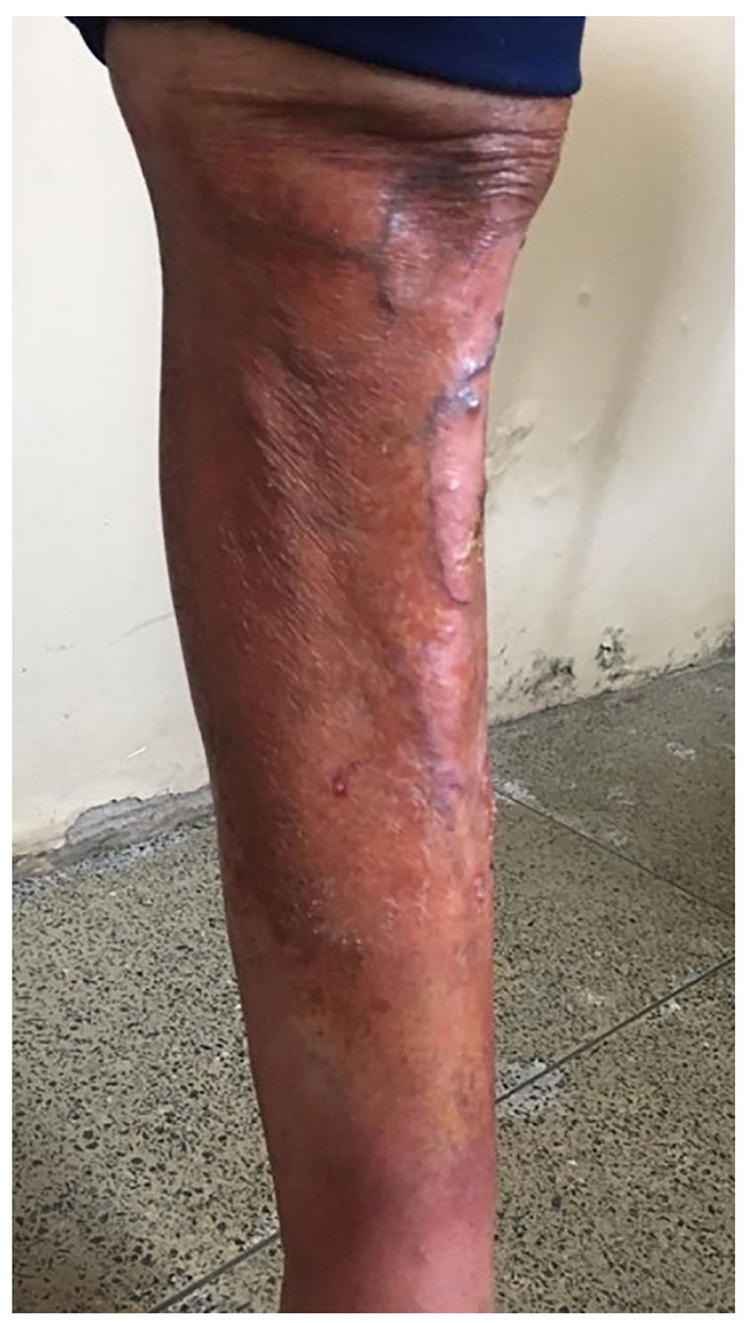
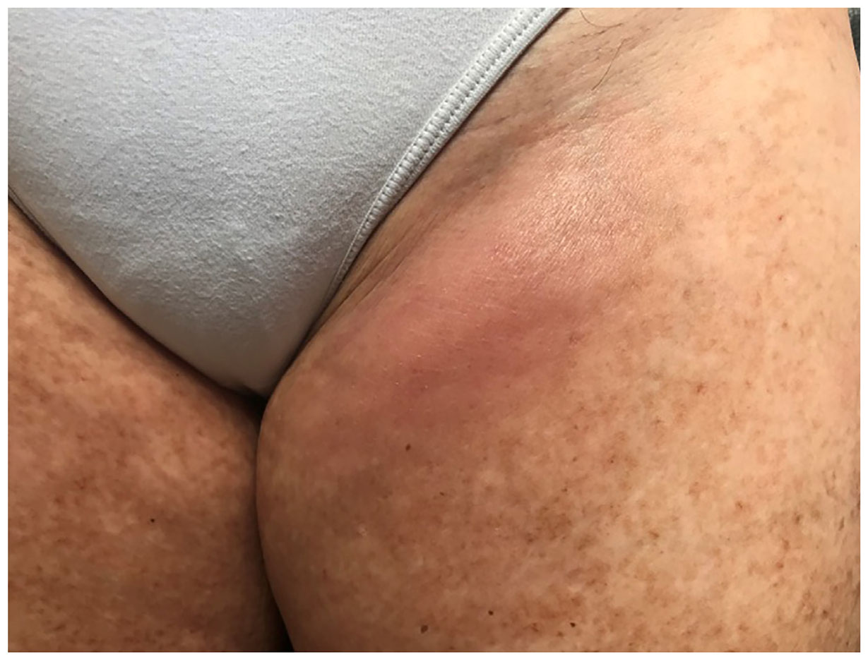
Photo: Brena Andrade de Lima Lobato.
Figure 2 - Hard lump under erythematous skin in the left inguinal region
DISCUSSION
The annual incidence of CTCL is extremely rare, of 0.77/100,000 individuals, and MF, the most common type of this lymphoma, has an approximate incidence of 0.41/100,000 individuals2.
MF may bear considerable resemblance to several benign inflammatory dermatoses, such as eczema, psoriasis, and pityriasis lichenoides chronica, and its classic histological features may be absent even in multiple skin biopsies, especially in their early stages, as observed in this case. Therefore, late diagnosis can result in the evolution and worsening of the patient's condition, including impairment in their quality of life, the need for more aggressive treatments, the occurrence of sequelae, and a possible iatrogenesis resulting from previous exams and treatments3,4,5,8. This issue was observed in the present case report, as, in six years, the patient had been evaluated by six physicians from different specialties until she was referred to the UEPA dermatology service and received a definitive diagnosis. This delay resulted in the worsening of the patient's condition and therapeutic difficulties, in addition to a significant waste of time and financial resources. The diagnostic delay observed was even greater than that found in studies conducted by Skov and Gniadecki9 and Scarisbrick et al.10, in which the meantime of diagnosis was two years and three months and three years, respectively. These points reinforce the understanding of MF as a public health problem, and the importance of a trained medical professional to distinguish cases to avoid further complications.
The disease staging was defined as T2NxM0B0 by the tumor-node-metastasis (TNM) staging system method, adapted to the specific parameters of the CTCL11. Thus, treatment with methotrexate 15 mg and folic acid 5 mg, in a single dose per week, associated with phototherapy with PUVA was carried out, and no adverse effects were observed. Treatment with PUVA phototherapy has benefit in MF remission, penetrating deeply into the dermis and can be used with systemic agents in severe cases7,12,13. Methotrexate, a folic acid analogue that belongs to the group of antimetabolic drugs, has proven benefits even at low doses; however, studies showing its use on a large scale are still scarce14,15. Furthermore, the association of methorexate with phototherapy showed promising results in the present case, reinforcing the possibility of its use as a therapeutic choice and subject of future studies.
CONCLUSION
MF is a lymphoproliferative cutaneous disease that is difficult to diagnose, especially in its early stages. This case report highlights the impacts of delayed diagnosis and the difficulty of medical professionals from different specialties in establishing this diagnosis, factors that impact the disease progression, worsen the prognosis, and may generate sequelae after treatment. Additionally, it shows the waste of time and financial resources of the patient, which affects their quality of life. MF is a major challenge for public health, especially in training and qualification of medical professionals.
ETHICAL ASPECTS
This case report was conducted during the project "Fototerapia em doenças linfoproliferativas e inflamatórias associadas ao HTLV", which was approved by the Research Ethics Committee of the UEPA, under No. 1.582.819, in June 9, 2016, in accordance with the precepts of the Declaration of Helsinki and the Nuremberg Code, respecting the standards of research involving human beings, Resolution No. 466/12 of the National Health Council.
REFERENCES
1 Willemze R, Jaffe ES, Burg G, Cerroni L, Berti E, Swerdlow SH, et al. WHO-EORTC classification for cutaneous lymphomas. Blood. 2005 May;105(10):3768-85. [ Links ]
2 Bradford PT, Devesa SS, Anderson WF, Toro JR. Cutaneous lymphoma incidence patterns in the United States: a population-based study of 3884 cases. Blood. 2009 May;113(21):5064-73. [ Links ]
3 Jawed SI, Myskowski PL, Horwitz S, Moskowitz A, Querfeld C. Primary cutaneous T-cell lymphoma (mycosis fungoides and Sézary syndrome): part I. Diagnosis: clinical and histopathologic features and new molecular and biologic markers. J Am Acad Dermatol. 2014 Feb;70(2):205.e1-16. [ Links ]
4 Foss FM, Girardi M. Mycosis fungoides and Sezary syndrome. Hematol Oncol Clin North Am. 2017 Apr;31(2):297-315. [ Links ]
5 Larocca C, Kupper T. Mycosis fungoides and Sézary syndrome: an update. Hematol Oncol Clin North Am. 2019 Feb;33(1):103-120. [ Links ]
6 Whittaker S, Hoppe R, Prince HM. How I treat mycosis fungoides and Sézary syndrome. Blood. 2016 Jun;127(25):3142-53. [ Links ]
7 Lovgren ML, Scarisbrick JJ. Update on skin directed therapies in mycosis fungoides. Chin Clin Oncol. 2019 Feb;8(1):7. [ Links ]
8 Jawed SI, Myskowski PL, Horwitz S, Moskowitz A, Querfeld C. Primary cutaneous T-cell lymphoma (mycosis fungoides and Sézary syndrome): part II. Prognosis, management, and future directions. J Am Acad Dermatol. 2014 Feb;70(2):223.e1-17. [ Links ]
9 Skov AG, Gniadecki R. Delay in the histopathologic diagnosis of mycosis fungoides. Acta Derm Venereol. 2015 Apr;95(4):472-5. [ Links ]
10 Scarisbrick JJ, Quaglino P, Prince HM, Papadavid E, Hodak E, Bagot M, et al. The PROCLIPI international registry of early-stage mycosis fungoides identifies substantial diagnostic delay in most patients. Br J Dermatol. 2019 Aug;181(2):350-7. [ Links ]
11 Olsen E, Vonderheid E, Pimpinelli N, Willemze R, Kim Y, Knobler R, et al. Revisions to the staging and classification of mycosis fungoides and Sezary syndrome: a proposal of the International Society for Cutaneous Lymphomas (ISCL) and the cutaneous lymphoma task force of the European Organization of Research and Treatment of Cancer (EORTC). Blood. 2007 Sep;110(6):1713-22. [ Links ]
12 Trautinger F. Phototherapy of cutaneous T-cell lymphomas. Photochem Photobiol Sci. 2018 Dec;17(12):1904-12. [ Links ]
13 Olsen EA, Hodak E, Anderson T, Carter JB, Henderson M, Cooper K, et al. Guidelines for phototherapy of mycosis fungoides and Sézary syndrome: a consensus statement of the United States Cutaneous Lymphoma Consortium. J Am Acad Dermatol. 2016 Jan;74(1): 27-58. [ Links ]
14 Olek-Hrab K, Maj J, Chmielowska E, Jankowska-Konsur A, Olszewska B, Kręcisz B, et al. Methotrexate in the treatment of mycosis fungoides - a multicenter observational study in 79 patients. Eur Rev Med Pharmacol Sci. 2018 Jun;22(11):3586-94. [ Links ]
15 Photiou L, van der Weyden C, McCormack C, Prince HM. Systemic treatment options for advanced-stage mycosis fungoides and Sézary syndrome. Curr Oncol Rep. 2018 Mar;20(4):32. [ Links ]
Received: October 22, 2020; Accepted: June 01, 2021
 Este é um artigo publicado em acesso aberto sob uma licença Creative Commons
