<?xml version="1.0" encoding="ISO-8859-1"?><article xmlns:mml="http://www.w3.org/1998/Math/MathML" xmlns:xlink="http://www.w3.org/1999/xlink" xmlns:xsi="http://www.w3.org/2001/XMLSchema-instance">
<front>
<journal-meta>
<journal-id>0101-5907</journal-id>
<journal-title><![CDATA[Revista Paraense de Medicina]]></journal-title>
<abbrev-journal-title><![CDATA[Rev. Para. Med.]]></abbrev-journal-title>
<issn>0101-5907</issn>
<publisher>
<publisher-name><![CDATA[Fundação Santa Casa de Misericórdia do Pará]]></publisher-name>
</publisher>
</journal-meta>
<article-meta>
<article-id>S0101-59072007000400002</article-id>
<title-group>
<article-title xml:lang="en"><![CDATA[Evaluation of APRI test as a liver fibrosis marker]]></article-title>
<article-title xml:lang="pt"><![CDATA[Avaliação do modelo APRI como marcador de fibrose hepática]]></article-title>
</title-group>
<contrib-group>
<contrib contrib-type="author">
<name>
<surname><![CDATA[Amaral]]></surname>
<given-names><![CDATA[Ivanete do Socorro Abraçado]]></given-names>
</name>
<xref ref-type="aff" rid="A01"/>
</contrib>
<contrib contrib-type="author">
<name>
<surname><![CDATA[Dias]]></surname>
<given-names><![CDATA[Marina Pinto Franco]]></given-names>
</name>
<xref ref-type="aff" rid="A02"/>
</contrib>
<contrib contrib-type="author">
<name>
<surname><![CDATA[Fernandes]]></surname>
<given-names><![CDATA[Clariana Casali Rodrigues]]></given-names>
</name>
<xref ref-type="aff" rid="A02"/>
</contrib>
<contrib contrib-type="author">
<name>
<surname><![CDATA[Moia]]></surname>
<given-names><![CDATA[Lizomar de Jesus Maués Pereira]]></given-names>
</name>
<xref ref-type="aff" rid="A03"/>
</contrib>
<contrib contrib-type="author">
<name>
<surname><![CDATA[Miranda]]></surname>
<given-names><![CDATA[Esther Castello Branco Mello]]></given-names>
</name>
<xref ref-type="aff" rid="A01"/>
</contrib>
<contrib contrib-type="author">
<name>
<surname><![CDATA[Demachki]]></surname>
<given-names><![CDATA[Sâmia]]></given-names>
</name>
<xref ref-type="aff" rid="A03"/>
</contrib>
</contrib-group>
<aff id="A01">
<institution><![CDATA[,State University of Pará  ]]></institution>
<addr-line><![CDATA[ ]]></addr-line>
</aff>
<aff id="A02">
<institution><![CDATA[,State University of Pará  ]]></institution>
<addr-line><![CDATA[ ]]></addr-line>
</aff>
<aff id="A03">
<institution><![CDATA[,Federal University of Pará  ]]></institution>
<addr-line><![CDATA[ ]]></addr-line>
</aff>
<pub-date pub-type="pub">
<day>00</day>
<month>12</month>
<year>2007</year>
</pub-date>
<pub-date pub-type="epub">
<day>00</day>
<month>12</month>
<year>2007</year>
</pub-date>
<volume>21</volume>
<numero>4</numero>
<fpage>7</fpage>
<lpage>13</lpage>
<copyright-statement/>
<copyright-year/>
<self-uri xlink:href="http://scielo.iec.gov.br/scielo.php?script=sci_arttext&amp;pid=S0101-59072007000400002&amp;lng=en&amp;nrm=iso"></self-uri><self-uri xlink:href="http://scielo.iec.gov.br/scielo.php?script=sci_abstract&amp;pid=S0101-59072007000400002&amp;lng=en&amp;nrm=iso"></self-uri><self-uri xlink:href="http://scielo.iec.gov.br/scielo.php?script=sci_pdf&amp;pid=S0101-59072007000400002&amp;lng=en&amp;nrm=iso"></self-uri><abstract abstract-type="short" xml:lang="en"><p><![CDATA[OBJECTIVE: to evaluate the performance of APRI test as an indirect marker of liver fibrosis in patients of FSCMPA with chronic viral hepatitis. METHODS: laboratorial and histopathological (METAVIR) data of 102 patients were collected with the aim of comparing biopsy reports to test results, considering: F0-F1 as absent/ insignificant fibrosis and F2-F4 as presence of expressive fibrosis; F0-F3 and F4 as absence and presence of cirrhosis, respectively. The evaluation was based on the use of simultaneous cut-off points suggested by Wai et al. (2003) and also the development of single cut-off points, through ROC curves, for this sample. RESULTS: the specificities found through the original cut-off points were 96% and 87% for significant fibrosis and cirrhosis, respectively. A superior performance was detected for the diagnosis of expressive fibrosis (PPV 90%) and for the exclusion of liver cirrhosis (NPV 95%). The only advantages accomplished with the use of the single cutoff points defined for this study were the classification of all patients and an increase in sensibility, which do not justify their applicability. CONCLUSION: APRI test is able to confirm the existence of significant fibrosis and exclude cirrhosis, what makes it available, as an alternative, in clinical practice.]]></p></abstract>
<abstract abstract-type="short" xml:lang="pt"><p><![CDATA[OBJETIVO: avaliar o desempenho do teste APRI como marcador indireto de fibrose hepática em pacientes portadores de hepatites virais crônicas da FSCMPA. MÉTODO: foram avaliados dados laboratoriais e histopatológicos (METAVIR) de 102 pacientes a fim de comparar os resultados da biópsia com o cálculo do teste. Considerou-se: F0-F1 e F2-F4 como fibrose ausente/inexpressiva e presença de fibrose expressiva, respectivamente; F0-F3 e F4 como ausência e presença de cirrose, respectivamente. Utilizou-se os pontos de corte simultâneos sugeridos por Wai et al. (2003) e desenvolveu-se pontos de corte únicos, através de curvas ROC, para esta amostra. RESULTADOS: as especificidades encontradas com os cortes originais foram, para fibrose significativa e cirrose, 96% e 87%, respectivamente. Evidenciou-se melhor desempenho ao confirmar diagnóstico de fibrose expressiva (VPP 90%) e em excluir cirrose hepática (VPN 95%). As únicas vantagens obtidas com os pontos definidos neste estudo foram a classificação de 100% dos pacientes e aumento da sensibilidade, o que não justifica sua aplicabilidade. CONCLUSÃO: o teste APRI é capaz de afirmar a existência de fibrose importante e de excluir o diagnóstico de cirrose, o que viabiliza seu uso, como alternativa, na prática clínica.]]></p></abstract>
<kwd-group>
<kwd lng="en"><![CDATA[APRI]]></kwd>
<kwd lng="en"><![CDATA[liver fibrosis]]></kwd>
<kwd lng="en"><![CDATA[evaluation]]></kwd>
<kwd lng="pt"><![CDATA[APRI]]></kwd>
<kwd lng="pt"><![CDATA[fibrose hepática]]></kwd>
<kwd lng="pt"><![CDATA[avaliação]]></kwd>
</kwd-group>
</article-meta>
</front><body><![CDATA[ <p align="right"><font size="2" face="verdana"><b><a name="topo"></a>ARTIGO ORIGINAL</b></font></p>     <p>&nbsp;</p>     <p><font size="4" face="verdana"><b>Evaluation of APRI test as a liver fibrosis    marker<sup><a href="#n1"><font size="3">1</font></a><a name="sup1"></a></sup></b></font></p>     <p>&nbsp;</p>     <p><font size="3" face="verdana"><b>Avalia&ccedil;&atilde;o do modelo APRI como    marcador de fibrose hep&aacute;tica<sup><a href="#n1">1</a></sup></b></font></p>     <p>&nbsp;</p>     <p>&nbsp;</p>     <p><font size="2" face="verdana"><b>Ivanete do Socorro Abra&ccedil;ado Amaral<sup>I</sup>;    Marina Pinto Franco Dias<sup>II</sup>; Clariana Casali Rodrigues Fernandes<sup>II</sup>;    Lizomar de Jesus Mau&eacute;s Pereira Moia<sup>III</sup>; Esther Castello Branco    Mello Miranda<sup>I</sup>; S&acirc;mia Demachki<sup>III</sup></b></font></p>     <p><font size="2" face="verdana"><sup>I</sup>Hepathologist and Professor of Adults'    Health (General Clinics) of State University of Par&aacute;    <br>   <sup>II</sup>Medicine 5<sup>th</sup>-year students of State University of Par&aacute;    ]]></body>
<body><![CDATA[<br>   <sup>III</sup>Hepathologist of Pathological Anatomy Departament of Federal University    of Par&aacute;</font></p>     <p><font size="2" face="verdana"><a href="#endereco">Correspondence </a></font></p>     <p>&nbsp;</p>     <p>&nbsp;</p> <hr size="1" noshade>     <p><font size="2" face="verdana"><b>SUMMARY</b></font></p>     <p><font size="2" face="verdana"><b>OBJECTIVE</b>: to evaluate the performance    of APRI test as an indirect marker of liver fibrosis in patients of FSCMPA with    chronic viral hepatitis.    <br>   <b>METHODS</b>: laboratorial and histopathological (METAVIR) data of 102 patients    were collected with the aim of comparing biopsy reports to test results, considering:    F0-F1 as absent/ insignificant fibrosis and F2-F4 as presence of expressive    fibrosis; F0-F3 and F4 as absence and presence of cirrhosis, respectively. The    evaluation was based on the use of simultaneous cut-off points suggested by    Wai et al. (2003) and also the development of single cut-off points, through    ROC curves, for this sample.    <br>   <b>RESULTS</b>: the specificities found through the original cut-off points    were 96% and 87% for significant fibrosis and cirrhosis, respectively. A superior    performance was detected for the diagnosis of expressive fibrosis (PPV 90%)    and for the exclusion of liver cirrhosis (NPV 95%). The only advantages accomplished    with the use of the single cutoff points defined for this study were the classification    of all patients and an increase in sensibility, which do not justify their applicability.    <br>   <b>CONCLUSION</b>: APRI test is able to confirm the existence of significant    fibrosis and exclude cirrhosis, what makes it available, as an alternative,    in clinical practice.</font></p>     <p><font size="2" face="verdana"><b>KEYWORDS</b>: APRI, liver fibrosis, evaluation.</font></p> <hr size="1" noshade>     ]]></body>
<body><![CDATA[<p><font size="2" face="verdana"><b>RESUMO</b></font></p>     <p><font size="2" face="verdana"><b><i>OBJETIVO:</i></b><i> avaliar o desempenho    do teste APRI como marcador indireto de fibrose hep&aacute;tica em pacientes    portadores de hepatites virais cr&ocirc;nicas da FSCMPA.    <br>   <b>M&Eacute;TODO:</b> foram avaliados dados laboratoriais e histopatol&oacute;gicos    (METAVIR) de 102 pacientes a fim de comparar os resultados da bi&oacute;psia    com o c&aacute;lculo do teste. Considerou-se: F0-F1 e F2-F4 como fibrose ausente/inexpressiva    e presen&ccedil;a de fibrose expressiva, respectivamente; F0-F3 e F4 como aus&ecirc;ncia    e presen&ccedil;a de cirrose, respectivamente. Utilizou-se os pontos de corte    simult&acirc;neos sugeridos por Wai et al. (2003) e desenvolveu-se pontos de    corte &uacute;nicos, atrav&eacute;s de curvas ROC, para esta amostra.    <br>   <b>RESULTADOS:</b> as especificidades encontradas com os cortes originais foram,    para fibrose significativa e cirrose, 96% e 87%, respectivamente. Evidenciou-se    melhor desempenho ao confirmar diagn&oacute;stico de fibrose expressiva (VPP    90%) e em excluir cirrose hep&aacute;tica (VPN 95%). As &uacute;nicas vantagens    obtidas com os pontos definidos neste estudo foram a classifica&ccedil;&atilde;o    de 100% dos pacientes e aumento da sensibilidade, o que n&atilde;o justifica    sua aplicabilidade.    <br>   <b>CONCLUS&Atilde;O:</b> o teste APRI &eacute; capaz de afirmar a exist&ecirc;ncia    de fibrose importante e de excluir o diagn&oacute;stico de cirrose, o que viabiliza    seu uso, como alternativa, na pr&aacute;tica cl&iacute;nica.</i></font></p>     <p><font size="2" face="verdana"><b>DESCRITORES</b>: APRI, fibrose hep&aacute;tica,    avalia&ccedil;&atilde;o.</font></p> <hr size="1" noshade>     <p>&nbsp;</p>     <p>&nbsp;</p>     <p><font size="3" face="verdana"><b>INTRODUCTION</b></font></p>     <p><font size="2" face="verdana">Liver fibrosis is a pathological process due    to   persistent and/or frequent hepatic damage. It is the   result of the inflammatory response or the direct toxic   action of a certain nocive agent, with posterior   parenchymal regeneration<sup>1,2</sup>.</font></p>     ]]></body>
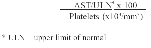
<body><![CDATA[<p><font size="2" face="verdana">The fibrosis degree is related, usually, to the   pathology severity. Hence, its evaluation is essential   for proper patients' management and follow-up.   Therefore, the exam confiability used to quantify   hepatic lesion is a vital feature<sup>1,3</sup>.</font></p>     <p><font size="2" face="verdana">Percutaneous liver biopsy is, nowadays, the gold   standard method for liver diseases evaluation and   evolutive analysis<sup>4,5</sup>.It is considered to be a safe procedure, however it is    not free of complications.   Besides, there are also limitations, related, mainly, to   contraindications, risks to the patient, technique, high   costs and interpretative inter/intraobserver variation   <sup>6,7,8,9,10</sup>.</font></p>     <p><font size="2" face="verdana">For those reasons, routine laboratory tests,   available in most health centers, have been used in   formulas in order to develop indexes that could obtain   biopsy-like results, i.e., being able to reflect   metabolical, morphological and functional changes   according to disease progression<sup>11</sup>.</font></p>     <p><font size="2" face="verdana">The AST-to-platelet ratio index (APRI), created    by Wai et al.<sup>3</sup> in 2003 is one of those tests. It is applied according    to the following formula:</font></p>     <p align="center"><font size="2" face="verdana"><img src="/img/revistas/rpm/v21n4/4a02for1.gif" border="0"></font></p>     <p><font size="2" face="verdana">This research attempts to demonstrate liver fibrosis    predictive ability of APRI test in patients with viral chronic hepatitis from    the Liver Group of Funda&ccedil;&atilde;o Santa Casa de Miseric&oacute;rdia    do Par&aacute; (FSCMPA) Hospital.</font></p>     <p>&nbsp;</p>     <p><font size="3" face="verdana"><b>OBJECTIVE</b></font></p>     <p><font size="2" face="verdana">To evaluate APRI test as a liver fibrosis marker    in viral chronic hepatitis, using biochemical, hematological and histopathological    data of patients registered at the Liver Group of FSCMPA Hospital.</font></p>     <p>&nbsp;</p>     ]]></body>
<body><![CDATA[<p><b><font size="3" face="verdana">METHODS</font></b></p>     <p><font size="2" face="verdana">This study was a retrospective analysis previously    approved by FSCMPA's Research Ethics Committee. Laboratorial and histopathological    data of 102 patients with chronic viral hepatitis were evaluated with the aim    of comparing biopsy reports to APRI results. Patients who had been through hepatitis    specific treatment were excluded, as well as those which laboratorial and histopathological    data were more than four months apart. The METAVIR grading was used, considering:    F0-F1 as absent/insignificant fibrosis and F2-F4 as presence of significant    fibrosis; F0-F3 and F4 as absence and presence of cirrhosis. Descriptive statistics    and correlation tests were elaborated by BioStat 4.0 software (P&quot;0,05=significance).    The evaluation of APRI was based on the use of simultaneous cut-off points suggested    by Wai et al. (2003)<sup>3</sup> and also the development, for this sample,    of single cut-off points through receiver operating characteristic (ROC) curves    constructed by MedCalc 9.3.7 software.</font></p>     <p>&nbsp;</p>     <p><font size="3" face="verdana"><b>RESULTS</b></font></p>     <p>&nbsp;</p>     <p align="center"><font size="2" face="verdana"><img src="/img/revistas/rpm/v21n4/4a02t1.gif" border="0"></font></p>     <p>&nbsp;</p>     <p>&nbsp;</p>     <p align="center"><font size="2" face="verdana"><img src="/img/revistas/rpm/v21n4/4a02f1.gif" border="0"></font></p>     <p>&nbsp;</p>     ]]></body>
<body><![CDATA[<p>&nbsp;</p>     <p align="center"><font size="2" face="verdana"><img src="/img/revistas/rpm/v21n4/4a02t2.gif" border="0"></font></p>     <p>&nbsp;</p>     <p>&nbsp;</p>     <p align="center"><font size="2" face="verdana"><img src="/img/revistas/rpm/v21n4/4a02t3.gif" border="0"></font></p>     <p>&nbsp;</p>     <p>&nbsp;</p>     <p align="center"><font size="2" face="verdana"><img src="/img/revistas/rpm/v21n4/4a02t4.gif" border="0"></font></p>     <p>&nbsp;</p>     <p>&nbsp;</p>     ]]></body>
<body><![CDATA[<p align="center"><font size="2" face="verdana"><img src="/img/revistas/rpm/v21n4/4a02f2.gif" border="0"></font></p>     <p>&nbsp;</p>     <p>&nbsp;</p>     <p align="center"><font size="2" face="verdana"><img src="/img/revistas/rpm/v21n4/4a02f3.gif" border="0"></font></p>     <p>&nbsp;</p>     <p>&nbsp;</p>     <p align="center"><font size="2" face="verdana"><img src="/img/revistas/rpm/v21n4/4a02f4.gif" border="0"></font></p>     <p>&nbsp;</p>     <p>&nbsp;</p>     <p align="center"><font size="2" face="verdana"><img src="/img/revistas/rpm/v21n4/4a02t5.gif" border="0"></font></p>     ]]></body>
<body><![CDATA[<p>&nbsp;</p>     <p>&nbsp;</p>     <p align="center"><font size="2" face="verdana"><img src="/img/revistas/rpm/v21n4/4a02t6.gif" border="0"></font></p>     <p>&nbsp;</p>     <p>&nbsp;</p>     <p align="center"><font size="2" face="verdana"><img src="/img/revistas/rpm/v21n4/4a02f5.gif" border="0"></font></p>     <p>&nbsp;</p>     <p>&nbsp;</p>     <p align="center"><font size="2" face="verdana"><img src="/img/revistas/rpm/v21n4/4a02f6.gif" border="0"></font></p>     <p>&nbsp;</p>     ]]></body>
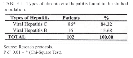
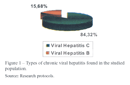
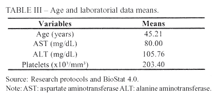
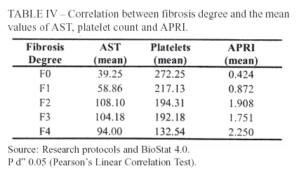
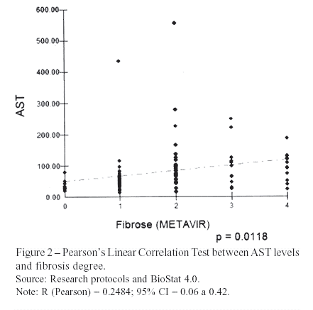
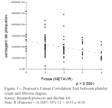
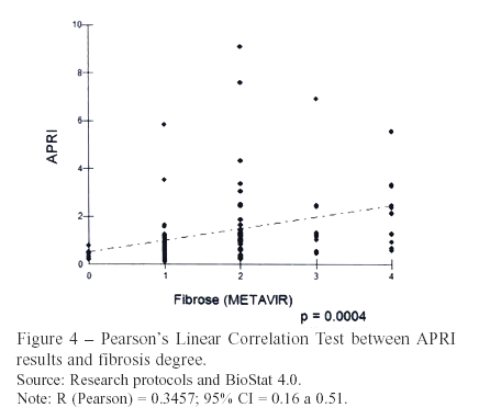
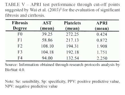
<body><![CDATA[<p>&nbsp;</p>     <p><font size="3" face="verdana"><b>DISCUSSION</b></font></p>     <p><font size="2" face="verdana">Models based on simple laboratorial tests have   been developed with the objective of validating   noninvasive methods that could, eventually, substitute   liver biopsy<sup>1</sup>. This research studied patients with   chronic viral hepatitis, being hepatitis C the most   prevalent one (84%). The remaining cases were all   type B, without any association with type D.</font></p>     <p><font size="2" face="verdana">The staging frequency of liver fibrosis after    biopsy,   according to METAVIR score, showed a prevalence   of F1 stage (42%), followed by F2 (28%), F3 and F4   (11% each)</font></p>     <p><font size="2" face="verdana">The studied patients presented an AST mean value    of 80mg/dL. Wai et al. (2006)<sup>7</sup>, evaluating only hepatitis B patients,    obtained means of 92 e 112mg/dL for training and validation sets, respectively.    The mean value of ALT was 106mg/dL, which is compatible to Iacobellis et al.    (2005),<sup>14</sup> result of 112mg/dL.</font></p>     <p><font size="2" face="verdana">Concerning the platelet count, the mean was 203,000/mm<sup>3</sup>.    This value is similar to most recent studies that aimed the validation of noninvasive    tests for liver fibrosis evaluation<sup>7,15,16,17</sup>.</font></p>     <p><font size="2" face="verdana">The elevation of AST according to fibrosis progression    is due to its clearance deficit and, also, increased mitochondrial damage<sup>18,19,20</sup>.    The patients of this research presented this trend, shown by Pearson's Linear    Correlation test &#8211; r = 0.25; <i>P</i> = 0.0118. However, the correlation    coefficient was less significant than the one achieved by the original study    of Wai et al. (2003)<sup>3</sup> &#8211; r = 0.50; <i>P</i> &lt; 0.01.</font></p>     <p><font size="2" face="verdana">Nonetheless, the mean values of AST according   to fibrosis degree increased until F2 and slightly   decreased at F3 and F4 stages. This feature could be   related to the smaller amount of parenchyma in precirrhotic   (F3) and cirrhotic patients, turning not possible   the damage of a great number of hepatocytes <sup>21</sup>.</font></p>     <p><font size="2" face="verdana">Thrombocytopenia is one of the most frequent    hematological disorders expected in patients with chronic liver disease, due,    primarily, to decreased hepatic thrombopoietin production and destruction of    platelets by splenomegaly<sup>11,22,23</sup>. Consequently, the evaluated patients    showed a progressive reduction of platelet count as fibrosis advanced. Pearson's    Linear Correlation (r = -0.37, <i>P</i> &lt;0.01) indicated this characteristic.    Wai et al. (2003)<sup>3</sup> obtained analogues results (r = -0.46; <i>P</i>    &lt; 0.01).</font></p>     <p><font size="2" face="verdana">Using the Pearson's Linear Correlation test for    APRI values and fibrosis stages (r = 0.35; <i>P</i> &lt;0.01), elevated values    of the index were associated to advanced fibrosis. Wai et al. (2003)<sup>3</sup>    accomplished a better coefficient (r = 0.60; <i>P</i> &lt;0.01).</font></p>     ]]></body>
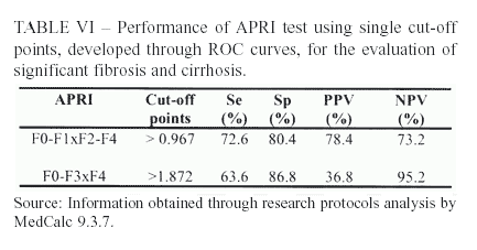
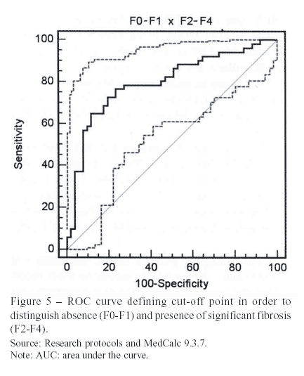
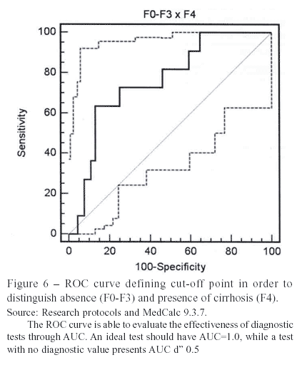
<body><![CDATA[<p><font size="2" face="verdana">Platelet count, therefore, showed the most   satisfactory correlation coefficient with fibrosis, when   compared to AST or APRI. Differently, Wai et al.   (2003)<sup>3</sup> developed APRI and reached the best   coefficient with the index.</font></p>     <p><font size="2" face="verdana">At Wai et al. (2003)<sup>3</sup> study, for the    determination of significant fibrosis, APRI d&quot; 0.5 showed sensibility (Se)    of 91% and &gt; 1.5 showed specificity (Sp) of 95%. The present survey reached    similar Sp (96.1%), while Se was lower (41.2%).</font></p>     <p><font size="2" face="verdana">The original study presented positive predictive   value (PPV) and negative predictive value (NPV) for   significant fibrosis of 88% and 85%, respectively. In   the actual study, only NPV achieved a non-satisfactory   percentage (60%).</font></p>     <p><font size="2" face="verdana">Wai et al. (2003)<sup>3</sup> also used APRI    to evaluate the presence of liver cirrhosis. For this quest, two distinct cut-off    points were defined: d&quot; 1.0 with 89% of Se and &gt; 2.0 with 93% of Sp.    The present research, after using the same points, obtained a modest Se (57.1%),    but a satisfactory Sp of 86.8%.</font></p>     <p><font size="2" face="verdana">Both studies, the present one and Wai et al.   (2003)<sup>3</sup>, reached excellent NPV's for cirrhosis, with   levels superior to 90%. On the other hand, the PPV   shown by the actual survey was unsatisfactory (36.8%)   as the original study was a little superior (57%).</font></p>     <p><font size="2" face="verdana">The results of APRI evaluation after using the    preestablished   cut-off points of 2003, in spite of   presenting different values from the original study, reaffirm that the index    has a good capacity to confirm   the diagnosis of significant fibrosis, besides being   essential to highlight its competence to exclude   patients without cirrhosis.</font></p>     <p><font size="2" face="verdana">Aiming to classify 100% of patients, single cutoff   points were developed through ROC curves. The   point established for significant fibrosis was &gt; 0.967.</font></p>     <p><font size="2" face="verdana">The AUROC (area under the ROC curve), able to   evaluate the diagnostic efficiency of the test, was   0.704, which is only a modest value, not excellent.</font></p>     <p><font size="2" face="verdana">Another ROC curve was also constructed for the   evaluation of cirrhosis, finding a single point of &gt;   1.872. The AUROC was, again, modest (0.758).</font></p>     <p><font size="2" face="verdana">However, using single cut-off points, there was   no relevant variation in the main parameters, except   for a slight increase in sensibility.</font></p>     ]]></body>
<body><![CDATA[<p><font size="2" face="verdana">The comparison between the original study's    Se,   Sp, PPV and NPV and the ones found in the present   study displayed an inferior upshot of the latter in most   parameters, whether using the cut-off points of Wai   et al. (2003)<sup>3</sup> or the single points defined for the actual   sample. It can be justified by the fact that patients   with hepatitis B, C and co-morbidities (HIV and   alcoholism) were studied in a unique group.</font></p>     <p><font size="2" face="verdana">Wai et al. (2006)<sup>7</sup> demonstrated, in a population   of only hepatitis B patients, that APRI was not able to   evaluate the presence of significant fibrosis and   cirrhosis with the same effectiveness of the original   study in hepatitis C patients. In the study of 2006, the   AUROC for significant fibrosis was 0.63, and 0.64   for cirrhosis. However, in the study of 2003 the   AUROC's were admirable (0.88 and 0.94).</font></p>     <p><font size="2" face="verdana">Hence, the interval of until four months between   liver biopsies and laboratorial results dates, adopted   in the present study, might have led to a result bias, as   patients with hepatitis B usually go through   aminotransferases fluctuation<sup>7</sup>.</font></p>     <p><font size="2" face="verdana">Using the single cut-off points, there was no   superior performance whether for fibrosis or cirrhosis.   This evidence shows that they are not likely to be used   in clinical practice, as the original Wai et al. (2003)<sup>3</sup>   cut-off points are more effective.</font></p>     <p><font size="2" face="verdana">The APRI is, therefore, able to prove the presence    of significant fibrosis and the absence of cirrhosis in a great number of patients    with viral chronic hepatitis. The index, however, is inadequate to evaluate    distinct etiologies of liver diseases through the same cut-off point.</font></p>     <p>&nbsp;</p>     <p><font size="3" face="verdana"><b>CONCLUSION</b></font></p>     <p><font size="2" face="verdana">It was shown that APRI is able to predict the   presence of significant fibrosis and cirrhosis through   the simultaneous cut-off points of Wai et al. (2003)<sup>3</sup>.   Although modest sensibilities were achieved, the   specificities for expressive fibrosis and cirrhosis were   96% e 87%, respectively. A better performance was   evidenced to confirm significant fibrosis (PPV = 90%)   and to exclude liver cirrhosis (NPV = 95%).</font></p>     <p><font size="2" face="verdana">Through the construction of ROC curves for   significant fibrosis and cirrhosis, it was possible to   classify the entire population of 102 patients by the   following cut-off points: &gt; 0.967 and &gt; 1.872,   respectively. Better sensibilities were found, which   were not enough to justify the use of those points as   the other parameters did not follow this tendency.</font></p>     <p><font size="2" face="verdana">It's important to emphasize the need of    new prospective studies, in order to validate the applicability of APRI in habitual    clinical practice. But still, to evaluate significant fibrosis and the exclusion    of cirrhosis, the index can be used as an alternative, mostly in patients with    contraindications for liver biopsy execution.</font></p>     ]]></body>
<body><![CDATA[<p>&nbsp;</p>     <p><font size="3" face="verdana"> <b>REFER&Ecirc;NCIAS</b></font></p>     <!-- ref --><p><font size="2" face="verdana">1 - COUTO, OFM. Valida&ccedil;&atilde;o e compara&ccedil;&atilde;o    de testes laboratoriais simples como preditores de fibrose hep&aacute;tica em    portadores de hepatite C cr&ocirc;nica. Available at: <a href="http://dspace.lcc.ufmg.br/dspace/bitstream/1843/ECJS-73AGQL/1/Osvaldo%2B%20Fl%C3%A1vio%2Bde%2BMelo%2BCouto.pdf" target="_blank">http://dspace.lcc.ufmg.br/dspace/bitstream/1843/ECJS-73AGQL/1/Osvaldo+    Fl%C3%A1vio+de+Melo+Couto.pdf</a>. Accessed on: June 25<sup>th</sup>, 2007.</font><!-- ref --><p><font size="2" face="verdana">2 - LEE, KS. Hepatic fibrogenesis. <i>Korean    J. Gastroenterol</i>. 2006; 48:297-305.</font><!-- ref --><p><font size="2" face="verdana">3 - WAI, CT et al. A simple noninvasive index    can predict both significant fibrosis and cirrhosis in patients with chronic    hepatitis C. <i>Hepatol</i>. 2003; 38:518-26.</font><!-- ref --><p><font size="2" face="verdana">4 - AFDHAL, NH; NUNES, D. Evaluation of liver    fibrosis: a concise review. <i>Am. J. Gastroenterol</i>. 2004;99:1160-74.</font><!-- ref --><p><font size="2" face="verdana">5 - ISHAK, K et al. Histological grading and    staging of chronic hepatitis. <i>J. Hepatol</i>.1995;22:696-9.</font><!-- ref --><p><font size="2" face="verdana">6 - TONIUTTO, P et al. Role of AST to platelet    ratio index in the detection of liver fibrosis in patients with recurrent hepatitis    C after liver transplantation. <i>J. Gastroenterol. Hepatol</i>.1997;22:1904-8.</font><!-- ref --><p><font size="2" face="verdana">7 - AI, C.T. et al. Noninvasive model for predicting    histology in patients with chronic hepatitis B. <i>Liver Int</i>.2006;26:666-72.</font><!-- ref --><p><font size="2" face="verdana">8 - AL-MOHRI, H et al. Validation of a simple    model for predicting liver fibrosis in HIV/hepatits C virus-coinfected patients.    <i>HIV Med</i>.2005;6:375-8.</font><!-- ref --><p><font size="2" face="verdana">9 - THULUVATH, PJ et al. Noninvasive markers    of fibrosis for longitudinal assessment of fibrosis in chronic liver disease:    Are they ready for prime time? <i>Am. J. Gastroenterol</i>.2005;100:1981-3.</font><!-- ref --><p><font size="2" face="verdana">10- POYNARD, T et al. Biomarkers as non-invasive    assessment of hepatic fibrosis in chronic hepatitis C. J. <i>Gastroenterol.    Hepatol</i>.2004;19:236-45.</font><!-- ref --><p><font size="2" face="verdana">11- TESTA, R et al. Noninvasive ratio index to    evaluate fibrosis staging in chronic hepatitis C: role of platelet count/spleen    diameter ratio index. <i>J. Intern. Med.</i>2006;260:142-50.</font><!-- ref --><p><font size="2" face="verdana">12- KELLEHER, TB; AFDHAL, N. Assessment of liver    fibrosis in co-infected patients. <i>J. Hepatol.</i>2006;44:126-31.</font><!-- ref --><p><font size="2" face="verdana">13- PARISE, ER et al. Noninvasive serum markers    in the diagnosis of structural liver damage in chronic hepatitis C virus infection.    <i>Liver Int.</i>2006;26:1095-9.</font><!-- ref --><p><font size="2" face="verdana">14- IACOBELLIS, A et al. External validation    of biochemical indices for noninvasive evaluation of liver fibrosis in HCV chronic    hepatitis. <i>Am. J. Gastroenterol.</i>2005;100:868-73.</font><!-- ref --><p><font size="2" face="verdana"> 15- BOURLIERE, M et al. Validation and comparison    of indexes for fibrosis and cirrhosis prediction in chronic hepatitis C patients:    proposal for a pragmatic approach classification without liver biopsies. <i>J.    Viral Hept.</i>2006;13:659-70.</font><!-- ref --><p><font size="2" face="verdana">16- MAOR, Y et al. Non-invasive biomarkers of    liver fibrosis in haemophilia patients with hepatitis C: can you avoid liver    biopsy? <i>Haemophilia</i>. 2006;12:372-9.</font><!-- ref --><p><font size="2" face="verdana">17- KELLEHER, TB et al. Prediction of hepatic    fibrosis in HIV/HCV co-infected patients using serum fibrosis markers: the SHASTA    index. <i>J. Hepatol.</i>2005;43:78-84.</font><!-- ref --><p><font size="2" face="verdana">18- OKUDA, M et al. Mitochondrial injury, oxidative    stress, and antioxidant gene expression are induced by hepatitis C virus core    protein. <i>Gastroentol</i>.2002;122:366-75.</font><!-- ref --><p><font size="2" face="verdana">19- HORIUCHI, S; KAMIMOTO, Y; MORINO, Y Hepatic    clearance of rat liver aspartate aminotransferase isozymes: evidence for endocytotic    uptake via different binding sites on sinusoidal liver cells. <i>Hepatol</i>.1985;5:376-82    apud TONIUTTO, P et al. Role of AST to platelet ratio index in the detection    of liver fibrosis in patients with recurrent hepatitis C after liver transplantation.    <i>J. Gastroenterol. Hepatol</i>.2007;22:1904-8.</font><!-- ref --><p><font size="2" face="verdana">20- NALPAS, B et al. Serum activity of mitochondrial    aspartate aminotransferase: a sensitive marker of alcoholism with or without    alcoholic hepatitis. <i>Hepatol.</i>, v. 4, p. 431-434, 1984 apud WAI, CT et    al. Noninvasive model for predicting histology in patients with chronic hepatitis    B. <i>Liver Int</i>.2006;26:666-72.</font><!-- ref --><p><font size="2" face="verdana">21- AMARAL, ISA. Co-infec&ccedil;&atilde;o v&iacute;rus    da imunodefici&ecirc;ncia humana e v&iacute;rus da hepatite C (HIV/HCV): aspectos    epidemiol&oacute;gicos, cl&iacute;nicos e laboratoriais de uma popula&ccedil;&atilde;o    atendida em um servi&ccedil;o de hepatopatias na cidade de Bel&eacute;m-Par&aacute;.    &#091;THESIS&#8211;MASTERS&#093;. Bel&eacute;m (PA): Federal University of Par&aacute;    &#8211; Health Sciences Center; 2006.</font><!-- ref --><p><font size="2" face="verdana">22- ADINOLFI, LE et al. Hepatic fibrosis plays    a central role in the pathogenesis of thrombocytopenia in patients with chronic    viral hepatitis. Br. <i>J. Haematol</i>.2001;113:590-5.</font><!-- ref --><p><font size="2" face="verdana">23- ASTER, RH. Pooling of platelets in the spleen:    role in the pathogenesis of 'hypersplenic' thrombocytopenia. <i>J. Clin. Invest.</i>,    v.45, p. 645-657, 1966 apud TONIUTTO, P et al. Role of AST to platelet ratio    index in the detection of liver fibrosis in patients with recurrent hepatitis    C after liver transplantation. <i>J. Gastroenterol. Hepatol</i>.2007;22:1904-8.</font><p>&nbsp;</p>     <p>&nbsp;</p>     <p><a name="endereco"></a><a href="#topo"><img src="/img/revistas/rpm/v21n4/seta.gif" border="0"></a><font size="2" face="verdana"><b>Address    mail</b><strong>:</strong>    <br>   Clariana Casali Rodrigues Fernandes    <br>   Avenida Jos&eacute; Bonif&aacute;cio 902, apto 702. S&atilde;o Braz, Bel&eacute;m -PA.    ]]></body>
<body><![CDATA[<br>   CEP: 66063-010 - Brazil    <br>   Telephone: +55 91 3249-9145.    <br>   <i>email</i>:<a href="mailto:clarianacasali@gmail.com">clarianacasali@gmail.com</a></font></p>     <p><font size="2" face="verdana">Ivanete do Socorro Abra&ccedil;ado Amaral    <br>   Avenida Gentil Bittencourt 1990, apto 1102. S&atilde;o Braz, Bel&eacute;m &#8211;PA    <br>   CEP: 66063-090 - Brazil    <br>   Telephone: +55 91 3272-0626    <br>   <i>email</i>:<a href="mailto:ivaneteabracado@hotmail.com">ivaneteabracado@hotmail.com</a></font></p>     <p><font size="2" face="verdana"> Lizomar de Jesus Mau&eacute;s Pereira Moia    <br>   Travessa Bom Jardim 658. Jurunas, Bel&eacute;m &#8211; PA    ]]></body>
<body><![CDATA[<br>   CEP: 66025-180 - Brazil    <br>   Telephone: +55 91 8844-4844    <br>   <i>email</i>: <a href="mailto:lizmoia@hotmail.com">lizmoia@hotmail.com</a></font></p>     <p><font size="2" face="verdana">Marina Pinto Franco Dias    <br>   Avenida Conselheiro Furtado 2818, apto 7000. S&atilde;o Braz, Bel&eacute;m -PA.    <br>   CEP: 66063-060 - Brazil    <br>   Telephone: +55 91 3229-0347.    <br>   <i>email</i>:<a href="mailto:marinafdias@gmail.com">marinafdias@gmail.com</a></font></p>     <p><font size="2" face="verdana">Recebido em 07.08.2007    <br>   Aprovado em 12.12.2007</font></p>     ]]></body>
<body><![CDATA[<p>&nbsp;</p>     <p>&nbsp;</p>     <p><font size="2" face="verdana"><sup><a name="n1"></a><a href="#sup1">1</a></sup>Study    performed at Funda&ccedil;&atilde;o Santa Casa de Miseric&oacute;rdia do Par&aacute;    Hospital</font></p>      ]]></body><back>
<ref-list>
<ref id="B1">
<label>1</label><nlm-citation citation-type="">
<person-group person-group-type="author">
<name>
<surname><![CDATA[COUTO]]></surname>
<given-names><![CDATA[OFM]]></given-names>
</name>
</person-group>
<source><![CDATA[Validação e comparação de testes laboratoriais simples como preditores de fibrose hepática em portadores de hepatite C crônica]]></source>
<year>June</year>
<month> 2</month>
<day>5t</day>
</nlm-citation>
</ref>
<ref id="B2">
<label>2</label><nlm-citation citation-type="journal">
<person-group person-group-type="author">
<name>
<surname><![CDATA[LEE]]></surname>
<given-names><![CDATA[KS]]></given-names>
</name>
</person-group>
<article-title xml:lang="en"><![CDATA[Hepatic fibrogenesis]]></article-title>
<source><![CDATA[Korean J. Gastroenterol]]></source>
<year>2006</year>
<volume>48</volume>
<page-range>297-305</page-range></nlm-citation>
</ref>
<ref id="B3">
<label>3</label><nlm-citation citation-type="journal">
<person-group person-group-type="author">
<name>
<surname><![CDATA[WAI]]></surname>
<given-names><![CDATA[CT]]></given-names>
</name>
</person-group>
<article-title xml:lang="en"><![CDATA[A simple noninvasive index can predict both significant fibrosis and cirrhosis in patients with chronic hepatitis C]]></article-title>
<source><![CDATA[Hepatol]]></source>
<year>2003</year>
<volume>38</volume>
<page-range>518-26</page-range></nlm-citation>
</ref>
<ref id="B4">
<label>4</label><nlm-citation citation-type="journal">
<person-group person-group-type="author">
<name>
<surname><![CDATA[AFDHAL]]></surname>
<given-names><![CDATA[NH]]></given-names>
</name>
<name>
<surname><![CDATA[NUNES]]></surname>
<given-names><![CDATA[D]]></given-names>
</name>
</person-group>
<article-title xml:lang="en"><![CDATA[Evaluation of liver fibrosis: a concise review]]></article-title>
<source><![CDATA[Am. J. Gastroenterol]]></source>
<year>2004</year>
<volume>99</volume>
<page-range>1160-74</page-range></nlm-citation>
</ref>
<ref id="B5">
<label>5</label><nlm-citation citation-type="journal">
<person-group person-group-type="author">
<name>
<surname><![CDATA[ISHAK]]></surname>
<given-names><![CDATA[K]]></given-names>
</name>
</person-group>
<article-title xml:lang="en"><![CDATA[Histological grading and staging of chronic hepatitis]]></article-title>
<source><![CDATA[J. Hepatol]]></source>
<year>1995</year>
<volume>22</volume>
<page-range>696-9</page-range></nlm-citation>
</ref>
<ref id="B6">
<label>6</label><nlm-citation citation-type="journal">
<person-group person-group-type="author">
<name>
<surname><![CDATA[TONIUTTO]]></surname>
<given-names><![CDATA[P]]></given-names>
</name>
</person-group>
<article-title xml:lang="en"><![CDATA[Role of AST to platelet ratio index in the detection of liver fibrosis in patients with recurrent hepatitis C after liver transplantation]]></article-title>
<source><![CDATA[J. Gastroenterol. Hepatol]]></source>
<year>1997</year>
<volume>22</volume>
<page-range>1904-8</page-range></nlm-citation>
</ref>
<ref id="B7">
<label>7</label><nlm-citation citation-type="journal">
<person-group person-group-type="author">
<name>
<surname><![CDATA[AI]]></surname>
<given-names><![CDATA[C.T]]></given-names>
</name>
</person-group>
<article-title xml:lang="en"><![CDATA[Noninvasive model for predicting histology in patients with chronic hepatitis B]]></article-title>
<source><![CDATA[Liver Int]]></source>
<year>2006</year>
<volume>26</volume>
<page-range>666-72</page-range></nlm-citation>
</ref>
<ref id="B8">
<label>8</label><nlm-citation citation-type="journal">
<person-group person-group-type="author">
<name>
<surname><![CDATA[AL-MOHRI]]></surname>
<given-names><![CDATA[H]]></given-names>
</name>
</person-group>
<article-title xml:lang="en"><![CDATA[Validation of a simple model for predicting liver fibrosis in HIV/hepatits C virus-coinfected patients]]></article-title>
<source><![CDATA[HIV Med]]></source>
<year>2005</year>
<volume>6</volume>
<page-range>375-8</page-range></nlm-citation>
</ref>
<ref id="B9">
<label>9</label><nlm-citation citation-type="journal">
<person-group person-group-type="author">
<name>
<surname><![CDATA[THULUVATH]]></surname>
<given-names><![CDATA[PJ]]></given-names>
</name>
</person-group>
<article-title xml:lang="en"><![CDATA[Noninvasive markers of fibrosis for longitudinal assessment of fibrosis in chronic liver disease: Are they ready for prime time?]]></article-title>
<source><![CDATA[Am. J. Gastroenterol]]></source>
<year>2005</year>
<volume>100</volume>
<page-range>1981-3</page-range></nlm-citation>
</ref>
<ref id="B10">
<label>10</label><nlm-citation citation-type="journal">
<person-group person-group-type="author">
<name>
<surname><![CDATA[POYNARD]]></surname>
<given-names><![CDATA[T]]></given-names>
</name>
</person-group>
<article-title xml:lang="en"><![CDATA[Biomarkers as non-invasive assessment of hepatic fibrosis in chronic hepatitis C]]></article-title>
<source><![CDATA[J. Gastroenterol. Hepatol]]></source>
<year>2004</year>
<volume>19</volume>
<page-range>236-45</page-range></nlm-citation>
</ref>
<ref id="B11">
<label>11</label><nlm-citation citation-type="journal">
<person-group person-group-type="author">
<name>
<surname><![CDATA[TESTA]]></surname>
<given-names><![CDATA[R]]></given-names>
</name>
</person-group>
<article-title xml:lang="en"><![CDATA[Noninvasive ratio index to evaluate fibrosis staging in chronic hepatitis C: role of platelet count/spleen diameter ratio index]]></article-title>
<source><![CDATA[J. Intern. Med]]></source>
<year>2006</year>
<volume>260</volume>
<page-range>142-50</page-range></nlm-citation>
</ref>
<ref id="B12">
<label>12</label><nlm-citation citation-type="journal">
<person-group person-group-type="author">
<name>
<surname><![CDATA[KELLEHER]]></surname>
<given-names><![CDATA[TB]]></given-names>
</name>
<name>
<surname><![CDATA[AFDHAL]]></surname>
<given-names><![CDATA[N]]></given-names>
</name>
</person-group>
<article-title xml:lang="en"><![CDATA[Assessment of liver fibrosis in co-infected patients]]></article-title>
<source><![CDATA[J. Hepatol]]></source>
<year>2006</year>
<volume>44</volume>
<page-range>126-31</page-range></nlm-citation>
</ref>
<ref id="B13">
<label>13</label><nlm-citation citation-type="journal">
<person-group person-group-type="author">
<name>
<surname><![CDATA[PARISE]]></surname>
<given-names><![CDATA[ER]]></given-names>
</name>
</person-group>
<article-title xml:lang="en"><![CDATA[Noninvasive serum markers in the diagnosis of structural liver damage in chronic hepatitis C virus infection]]></article-title>
<source><![CDATA[Liver Int]]></source>
<year>2006</year>
<volume>26</volume>
<page-range>1095-9</page-range></nlm-citation>
</ref>
<ref id="B14">
<label>14</label><nlm-citation citation-type="journal">
<person-group person-group-type="author">
<name>
<surname><![CDATA[IACOBELLIS]]></surname>
<given-names><![CDATA[A]]></given-names>
</name>
</person-group>
<article-title xml:lang="en"><![CDATA[External validation of biochemical indices for noninvasive evaluation of liver fibrosis in HCV chronic hepatitis]]></article-title>
<source><![CDATA[Am. J. Gastroenterol]]></source>
<year>2005</year>
<volume>100</volume>
<page-range>868-73</page-range></nlm-citation>
</ref>
<ref id="B15">
<label>15</label><nlm-citation citation-type="journal">
<person-group person-group-type="author">
<name>
<surname><![CDATA[BOURLIERE]]></surname>
<given-names><![CDATA[M]]></given-names>
</name>
</person-group>
<article-title xml:lang="en"><![CDATA[Validation and comparison of indexes for fibrosis and cirrhosis prediction in chronic hepatitis C patients: proposal for a pragmatic approach classification without liver biopsies]]></article-title>
<source><![CDATA[J. Viral Hept]]></source>
<year>2006</year>
<volume>13</volume>
<page-range>659-70</page-range></nlm-citation>
</ref>
<ref id="B16">
<label>16</label><nlm-citation citation-type="journal">
<person-group person-group-type="author">
<name>
<surname><![CDATA[MAOR]]></surname>
<given-names><![CDATA[Y]]></given-names>
</name>
</person-group>
<article-title xml:lang="en"><![CDATA[Non-invasive biomarkers of liver fibrosis in haemophilia patients with hepatitis C: can you avoid liver biopsy?]]></article-title>
<source><![CDATA[Haemophilia]]></source>
<year>2006</year>
<volume>12</volume>
<page-range>372-9</page-range></nlm-citation>
</ref>
<ref id="B17">
<label>17</label><nlm-citation citation-type="journal">
<person-group person-group-type="author">
<name>
<surname><![CDATA[KELLEHER]]></surname>
<given-names><![CDATA[TB]]></given-names>
</name>
</person-group>
<article-title xml:lang="en"><![CDATA[Prediction of hepatic fibrosis in HIV/HCV co-infected patients using serum fibrosis markers: the SHASTA index]]></article-title>
<source><![CDATA[J. Hepatol]]></source>
<year>2005</year>
<volume>43</volume>
<page-range>78-84</page-range></nlm-citation>
</ref>
<ref id="B18">
<label>18</label><nlm-citation citation-type="journal">
<person-group person-group-type="author">
<name>
<surname><![CDATA[OKUDA]]></surname>
<given-names><![CDATA[M]]></given-names>
</name>
</person-group>
<article-title xml:lang="en"><![CDATA[Mitochondrial injury, oxidative stress, and antioxidant gene expression are induced by hepatitis C virus core protein]]></article-title>
<source><![CDATA[Gastroentol]]></source>
<year>2002</year>
<volume>122</volume>
<page-range>366-75</page-range></nlm-citation>
</ref>
<ref id="B19">
<label>19</label><nlm-citation citation-type="journal">
<person-group person-group-type="author">
<name>
<surname><![CDATA[HORIUCHI]]></surname>
<given-names><![CDATA[S]]></given-names>
</name>
<name>
<surname><![CDATA[KAMIMOTO]]></surname>
<given-names><![CDATA[Y]]></given-names>
</name>
<name>
<surname><![CDATA[MORINO]]></surname>
<given-names><![CDATA[Y]]></given-names>
</name>
</person-group>
<article-title xml:lang="en"><![CDATA[Hepatic clearance of rat liver aspartate aminotransferase isozymes: evidence for endocytotic uptake via different binding sites on sinusoidal liver cells. Hepatol.1985;5:376-82 apud TONIUTTO, P et al. Role of AST to platelet ratio index in the detection of liver fibrosis in patients with recurrent hepatitis C after liver transplantation]]></article-title>
<source><![CDATA[J. Gastroenterol. Hepatol]]></source>
<year>2007</year>
<volume>22</volume>
<page-range>1904-8</page-range></nlm-citation>
</ref>
<ref id="B20">
<label>20</label><nlm-citation citation-type="journal">
<person-group person-group-type="author">
<name>
<surname><![CDATA[NALPAS]]></surname>
<given-names><![CDATA[B]]></given-names>
</name>
</person-group>
<article-title xml:lang="en"><![CDATA[Serum activity of mitochondrial aspartate aminotransferase: a sensitive marker of alcoholism with or without alcoholic hepatitis. Hepatol., v. 4, p. 431-434, 1984 apud WAI, CT et al. Noninvasive model for predicting histology in patients with chronic hepatitis B]]></article-title>
<source><![CDATA[Liver Int]]></source>
<year>2006</year>
<volume>26</volume>
<page-range>666-72</page-range></nlm-citation>
</ref>
<ref id="B21">
<label>21</label><nlm-citation citation-type="">
<person-group person-group-type="author">
<name>
<surname><![CDATA[AMARAL]]></surname>
<given-names><![CDATA[ISA]]></given-names>
</name>
</person-group>
<source><![CDATA[Co-infecção vírus da imunodeficiência humana e vírus da hepatite C (HIV/HCV): aspectos epidemiológicos, clínicos e laboratoriais de uma população atendida em um serviço de hepatopatias na cidade de Belém-Pará]]></source>
<year></year>
</nlm-citation>
</ref>
<ref id="B22">
<label>22</label><nlm-citation citation-type="journal">
<person-group person-group-type="author">
<name>
<surname><![CDATA[ADINOLFI]]></surname>
<given-names><![CDATA[LE]]></given-names>
</name>
</person-group>
<article-title xml:lang="en"><![CDATA[Hepatic fibrosis plays a central role in the pathogenesis of thrombocytopenia in patients with chronic viral hepatitis]]></article-title>
<source><![CDATA[Br. J. Haematol]]></source>
<year>2001</year>
<volume>113</volume>
<page-range>590-5</page-range></nlm-citation>
</ref>
<ref id="B23">
<label>23</label><nlm-citation citation-type="journal">
<person-group person-group-type="author">
<name>
<surname><![CDATA[ASTER]]></surname>
<given-names><![CDATA[RH]]></given-names>
</name>
</person-group>
<article-title xml:lang="en"><![CDATA[Pooling of platelets in the spleen: role in the pathogenesis of 'hypersplenic' thrombocytopenia. J. Clin. Invest., v.45, p. 645-657, 1966 apud TONIUTTO, P et al. Role of AST to platelet ratio index in the detection of liver fibrosis in patients with recurrent hepatitis C after liver transplantation]]></article-title>
<source><![CDATA[J. Gastroenterol. Hepatol]]></source>
<year>2007</year>
<volume>22</volume>
<page-range>1904-8</page-range></nlm-citation>
</ref>
</ref-list>
</back>
</article>